Pharma Strategy Blog

Commentary on Pharma & Biotech Oncology / Hematology New Product Development

Posts from the ‘Basic Research’ category

Today’s Science Friday post looks at the identification of a potential new biomarker and possible strategies for expanding use of PARP inhibitors in patients most likely to respond to them as a way to validate the the approach prospectively.  This has important implications for future clinical trial designs with this class of drugs.

Photo Credit: Ben Sutherland via flickr

Photo Credit: Ben Sutherland via flickr

Regular readers will be very familiar at my rants against broad catch-all studies and phase III trials with targeted agents that do not have a biomarker or even a logical well defined subset of patients because it’s akin to blindfolding an archer, turning him around 360 degrees and then asking him to hit a bullseye 50 or 100 yards hence.

How can you hit a target you can’t see?

PARP inhibitors have had a bit of a chequered history after the initial excitement was later followed by a series of rather disappointing clinical trial results, which occurred for a multitude of reasons.  Several of the approaches sadly fell into the category of ‘Five things not to do in R&D’ as delineated in my recent tongue-firmly-in-cheek post on the topic.  I’ll leave interested and curious readers to ponder at length exactly which of the no-nos they managed to break!

That said, we do know a few things of relevance:

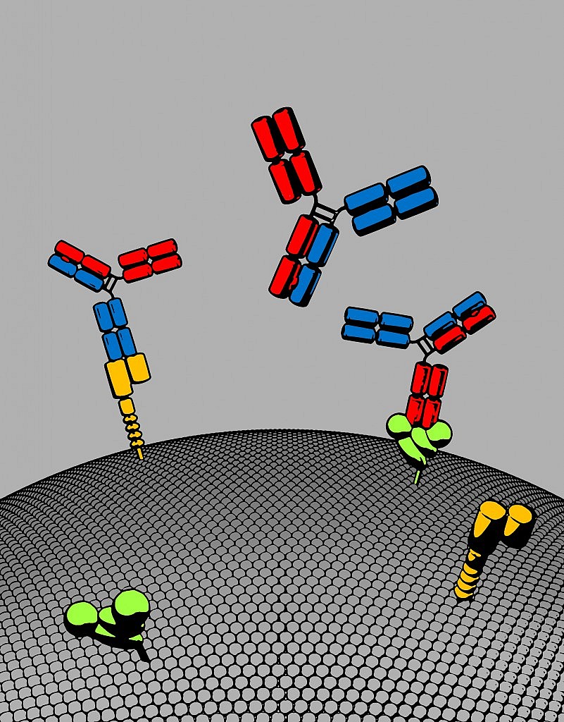
  1. PARP1/2 inhibitors have produced sustained anti-tumor responses in patients with germ-line BRCA gene mutations
  2. Biomarkers other than BRCA1 and BRCA2 are clearly needed to predict responses to PARP inhibitors
  3. Catch-all trials without segmenting for BRCA mutations and biomarkers are highly unlikely to be successful (recall the spectacular phase III failure in triple negative breast cancer with iniparib, for example)
  4. Next generation PARP inhibitors have focused on more clearly defined subset populations with greater success (and potency).

Chris Lord and Alan Ashworth’s lab have recently published some nice work (open access, see Refs below) that is worthy of discussion. Bajrami et al., (2013) decided to dive a little deeper and look at what genes and mutations might affect the responses to PARP inhibition:

“In hypothesizing that additional genetic determinants might direct use of these drugs, we conducted a genome-wide synthetic lethal screen for candidate olaparib sensitivity genes.  In support of this hypothesis, the set of identified genes included known determinants of olaparib sensitivity, such as BRCA1, RAD51 and Fanconi’s anemia susceptibility genes.”

The main reason behind this approach quickly becomes apparent – there is a huge need for it:

“One of the major issues in the clinical development of PARP1/2 inhibitors is the identification of biomarkers other than BRCA1 and BRCA2 gene mutations that predict a favourable response to therapy.”

While patients with germline BRCA mutations have a greater tendency to respond, they don’t account for all the responses that have been seen in the clinic.  What else is involved?

What did they find?

“Integration of the list of candidate sensitivity genes with data from tumor DNA sequencing studies identified CDK12 deficiency as a clinically relevant biomarker of PARP1/2 inhibitor sensitivity.”

In other words, when CDK12 was low, PARP sensitivity was high, making it a potential predictive biomarker for response to therapy with a PARP inhibitor.

What does this mean?

“The appearance of CDK12 in our olaparib sensitization gene list, alongside the CDK12 mutational data, suggested the possibility that loss of CDK12 function could sensitize tumor cells to PARP1/2 inhibitors and that loss of CDK12 function in HGSOVCa could be a predictive biomarker for response to this developmental class of agents.”

In other words, it would be useful to evaluate loss of CDK12 as a biomarker of response for PARP inhibitors in prospective clinical trials in breast and ovarian cancers, with and without germ-line BRCA mutations.

CDK12 is only one of nine genes known to be mutated in high grade serous ovarian cancer (HGS-OVCa), for example, so should patients be identified upfront who have loss of CDK12, then it may be enough to ensure response to a PARP inhibitor, irrespective of BRCA status.  Clearly this work is still very early, but it creates a smart and well argued rationale that can be tested in clinical trials.

Should the approach be validated, then it could well expand the utility of PARP inhibitors in the clinic based on a predictive biomarker.  I would be interested to see what happens not only with olaparib, but also the new generation of PARP inhibitors from Abbott (veliparib), Biomarin (BMN 673) and Clovis (rucaparib), to name a few who are conducting trials in breast or ovarian cancers.

If this works, it will be a thing of beauty.

References:

ResearchBlogging.orgBajrami I, Frankum JR, Konde A, Miller RE, Rehman FL, Brough R, Campbell J, Sims D, Rafiq R, Hooper S, Chen L, Kozarewa I, Assiotis I, Fenwick K, Natrajan R, Lord CJ, & Ashworth A (2014). Genome-wide Profiling of Genetic Synthetic Lethality Identifies CDK12 as a Novel Determinant of PARP1/2 Inhibitor Sensitivity. Cancer research, 74 (1), 287–97 PMID: 24240700

Photo Credit: Ben Sutherland

“RAF inhibitors (vemurafenib and dabrafenib) have profound clinical activity in patients with BRAF-mutant melanoma, but their therapeutic effects are limited by the emergence of drug resistance.”

Solit and Rosen (2014)

For today’s post on Science Fridays, I wanted to take a look at an overview paper, published in Cancer Discovery, from two researchers in the metastatic melanoma field who have been looking at multiple mechanisms of resistance.  It’s an important topic because while we have seen incremental improvements in outcomes for this disease, the 5-year survival rate is still rather poor with only 10–20% of metastatic patients still alive by then.  This is not to disparage the efforts of scientists, clinicians or companies working in this space, far from it, but there is is clearly a need for new therapies, strategies and combinations, given the high unmet medical need that exists.

We still have a long way to go in moving the survival needle dramatically.

It wasn’t until I searched for related blog posts to link to this one that I realised how much we have already covered on this topic! Regular readers will recall discussions here on PSB on various combinations such as:

  1. RAF + MEK inhibitors (downstream resistance)
  2. RAF + PI3K-AKT-mTOR inhibitors (cross resistance)
  3. RAF + CTLA–4 checkpoint inhibitors (anti-tumour immunity)

to name a few examples.

We have seen that adding a MEK inhibitor to dabrafenib e.g. trametinib can overcome resistance temporarily and add a few extra months before the resistance sets in again. Similarly for PI3K inhibitors tested to date. Adding ipilimumab, an anti-CTLA–4 checkpoint inhibitor held much promise, but the combination was abandoned with the emergence of unexpected liver toxicity.

Results thus far suggest that something else is acting as an escape route, thereby enabling the tumour to continue driving oncogenic addiction to BRAF.

The $64K questions are what is happening and what can we do about it?

We also need to remember that clinical research advances piecemeal based on evidence from preclinical reseach, so we see the logical evolution of BRAF monotherapy -> combos with downstream (MEK) or upstream (NRAS) targets in same pathway -> combos with diagonal (PI3K) pathways etc.

What Solit and Rosen have done is put a nice summary together of the state of play in this disease and the paper (see References below) is well worth reading.

Their main assertion is interesting, namely:

“The common feature of each of these mechanisms of resistance is that they result in activation of ERK signaling that is insensitive to the RAF inhibitor. Thus, RAF inhibitor resistance is often associated with maintenance of activation of the oncogene-driven pathway.”

Two recent papers are cited in support of this theory from Shi et al., (2014) and Allen et al., (2014) – see References below for additional background reading. Both studies used patient samples to look at clonal evolution and the genetic landscape in advanced melanoma. It’s actually quite amazing what unbiased exome sequencing can uncover at the molecular level, not least are the development of new mutations and other functional alterations.

The Shi et al., (2014) study was briefly summarised by Solit and Rosen:

“Multiple biopsies were obtained at different times or from disparate locations from several patients, and more than a single lesion in the ERK pathway was identifi ed in multiple patients typically within
different tumor biopsies.”

They went to note:

“A detailed phylogenetic analysis of multiple progressive lesions from a subset of these patients suggested branching evolution of tumors in which the development of genetic diversity was not linearly associated with time.”

Previously, a case report found distinct mechanisms of BRAF inhibitor resistance were present in two different progressing lesions from a single patient, so the work of Shi et al., (2014) is consistent with this finding. It blows my mind that different lesions in the same patient might behave completely differently though – imagine trying to devise an appropriate and effective clinical strategy in these cases?!

Allen et al’s (2014) work also involved whole exome sequencing (WES) from patient samples:

“WES was performed on paired pretreatment and progression samples collected from 45 patients, of whom 14 developed resistance soon after initiation of therapy (within 12 weeks). They also detected several resistance mechanisms that had been previously identified to confer RAF inhibitor resistance, including mutations in NRAS , MAP2K1, and NF1 and BRAF amplification.”

A third important study in this area from Wagle et al., (2014) adds to the weight of evidence with new mutations developing. Solit and Rosen continued the story:

“Consistent with the preclinical studies highlighted above demonstrating that MEK1 and MEK2
mutations can confer RAF and MEK inhibitor resistance, a MEK2 Q60P mutation was identifi ed in 1 of 5 patients studied. Of greater surprise to the investigators, one patient had a BRAF splice variant lacking exons 2–10 and a second patient had BRAF amplification.”

By now, you can see the sheer variety of changes and adaptations taking place in different studies around the world in some of the top melanoma labs. What do they have in common though?

“One hypothesis to explain this result is that increased abundance of the oncogenic driver (in this case BRAF) in response to prolonged drug treatment results in increased flux through the ERK pathway and restoration of ERK activity above the threshold required for inhibition of cell proliferation.”

The next challenge is to figure how we can approach better therapeutic index and shutting down of the pathways?

“The results suggest that the early adaptive response of BRAF -mutant cells to ERK pathway inhibition may promote the selection of resistant clones that harbor additional genomic events that
confer higher levels of RAF inhibitor resistance. The data also support combinatorial approaches that attenuate the adaptive response, including the addition of a PI3K or AKT inhibitor to the RAF and MEK (or ERK) inhibitor combination.”

The problem with this approach though, is that the neither the BRAF nor PI3K inhibitors have been able to reach or go beyond the single agent dosing schedules:

“As previous attempts to combine MAPK and PI3K pathway inhibitors have been limited by overlapping toxicities, upfront testing of intermittent treatment schedules should be considered.”

This is the also approach that Das Thakur suggested in her work presented at AACR last year, and subsequently published in Nature, to delay the development of resistance to vemurafenib.

I do think this one area where we may well see new trials evolve in advanced melanoma, so we will have to wait for new data before we can see if the strategy is successful at delaying the emergence of resistant clones. It is good to see the evolution of solid preclinical and translational evidence from patient biopsies helping to inform future clinical trial strategies.

In the meantime, the next major milestone I’m waiting for is on Roche/Genentech’s MEK inhibitor, cobimetinib (GDC–0973), which is due to report combination data with vemurafenib (continuous dosing) later this year. It will be interesting to see if this inhibits MEK more completely than trametinib and whether the combination has a better initial outcome than dabrafenib plus trametinib, which added about two to three months of extra survival over dabrafenib alone.

References:

ResearchBlogging.orgSolit DB, & Rosen N (2014). Towards a Unified Model of RAF Inhibitor Resistance. Cancer discovery, 4 (1), 27–30 PMID: 24402945

Shi H, Hugo W, Kong X, Hong A, Koya RC, Moriceau G, Chodon T, Guo R, Johnson DB, Dahlman KB, Kelley MC, Kefford RF, Chmielowski B, Glaspy JA, Sosman JA, van Baren N, Long GV, Ribas A, & Lo RS (2014). Acquired Resistance and Clonal Evolution in Melanoma during BRAF Inhibitor Therapy. Cancer discovery, 4 (1), 80–93 PMID: 24265155

Van Allen EM, Wagle N, Sucker A, Treacy DJ, Johannessen CM, Goetz EM, Place CS, Taylor-Weiner A, Whittaker S, Kryukov GV, Hodis E, Rosenberg M, McKenna A, Cibulskis K, Farlow D, Zimmer L, Hillen U, Gutzmer R, Goldinger SM, Ugurel S, Gogas HJ, Egberts F, Berking C, Trefzer U, Loquai C, Weide B, Hassel JC, Gabriel SB, Carter SL, Getz G, Garraway LA, Schadendorf D, & Dermatologic Cooperative Oncology Group of Germany (DeCOG) (2014). The Genetic Landscape of Clinical Resistance to RAF Inhibition in Metastatic Melanoma. Cancer discovery, 4 (1), 94–109 PMID: 24265153

Wagle N, Van Allen EM, Treacy DJ, Frederick DT, Cooper ZA, Taylor-Weiner A, Rosenberg M, Goetz EM, Sullivan RJ, Farlow DN, Friedrich DC, Anderka K, Perrin D, Johannessen CM, McKenna A, Cibulskis K, Kryukov G, Hodis E, Lawrence DP, Fisher S, Getz G, Gabriel SB, Carter SL, Flaherty KT, Wargo JA, & Garraway LA (2014). MAP Kinase Pathway Alterations in BRAF-Mutant Melanoma Patients with Acquired Resistance to Combined RAF/MEK Inhibition. Cancer discovery, 4 (1), 61–8 PMID: 24265154

Das Thakur M, Salangsang F, Landman AS, Sellers WR, Pryer NK, Levesque MP, Dummer R, McMahon M, & Stuart DD (2013). Modelling vemurafenib resistance in melanoma reveals a strategy to forestall drug resistance. Nature, 494 (7436), 251–5 PMID: 23302800

At AACR last year, one of the most revealing presentations was on metastatic melanoma, specifically, some elegant research by Meghna Das Thakur (NIBR) demonstrating that intermittent pulsing of vemurafenib (a BRAF V600E inhibitor) led to less resistance than inhibiting the target 24/7.

Many of us wondered whether such a pulsing approach would be useful for other tyrosine kinase inhibitors (TKIs).

Fast forward to this week.

CD current Jan 2014Neal Rosen’s lab at MSKCC has an interesting new paper out looking at the effects of pulse dosing with PI3K and ERK inhibition, since targeting both has long been suspected to be key in overcoming cross-resistance.

Recall that despite promising preclinical research, most of the early patient trials looking at targeting the PI3K-Akt-mTOR and RAS-RAF-MEK-ERK pathways in combination were, however, disappointing to say the least, both in terms of the side effect profile, and also with respect to clinical efficacy.

These results also applied to combinations with chemotherapy, which were added to either agent to try and induce cell death via apoptosis.

We know that the PI3K pathway is dysregulated in many cancers, so why have the combinations tried to date produced less than optimal results?

Well, Will et al., (2014) showed that:

  1. RAS-ERK pathway is a key downstream effector pathway of oncogenic PI3K
  2. ERK inhibition is required for apoptosis (cell death) to occur with a PI3K inhibitor
  3. It is important to coordinate downregulation of AKT and ERK since both are necessary for induction of apoptosis and antitumor activity
  4. Such an effect can be achieved with intermittent dosing, which will also likely decrease toxicity and allow administration of therapeutic doses

Ah so the same concept that Das explored in metastatic melanoma could also have potential with PI3K and MEK inhibition!

I find this approach fascinating because in the past, when I queried whether we needed to hit two targets maximally and continuously, rather than look at intermittent or minimally effective dosing (MED), industry people were up in arms and sent me more heated emails on this topic than anything else we’ve ever blogged about!

Meanwhile, Rosen himself hinted at this solution in a talk at the AACR Molecular Targets meeting in Boston last year and said a publication was underway to explain their findings. Generally, I don’t report on unpublished findings out of respect to the scientists and thus didn’t mention it in our extensive AACR Targets Coverage, but am delighted this is now a topic for more public discussion.

Part of the conundrum was articulated by Will et al., (2014) in their author manauscript (see below for the link under the Cancer Discovery Online First Section this month):

“Since mTOR and AKT inhibitors reactivate PI3K signaling, we asked whether PI3K inhibitors have more significant antitumor activity, perhaps by inhibiting other PI3K targets in addition to AKT/mTOR.

Selective PI3K and AKT inhibitors were compared in tumors with activation of PI3K pathway signaling in order to assess differences in the biochemical and biologic consequences of their inhibition. Both inhibitors effectively inhibited downstream targets of AKT, relieved feedback inhibition of growth factor receptors, and inhibited cell growth. However, in HER2-dependent breast cancers, PI3K inhibitors, but not AKT inhibitors, caused the rapid induction of a significant degree of apoptosis.

We find that, whereas AKT inhibitors inhibit AKT/mTOR and activate ERK signaling, PI3K inhibitors inhibit both. They cause durable inhibition of AKT signaling but also transient inhibition of RAS activation and ERK signaling, both of which are required for induction of apoptosis. Moreover, induction of apoptosis by an AKT inhibitor is significantly enhanced when combined with a MEK inhibitor.

Our results show that PI3K is upstream of wild type RAS as well as AKT/mTOR, and this causes the therapeutic consequences of PI3K inhibition to be significantly greater than those of AKT inhibition.”

A number of different inhibitors of PI3K, AKT, mTOR and MEK were explored in this research, so the results are not limited to one or two.

One important question that the group sought to address the inhibition issue:

“PI3K inhibitors cause rapid inhibition of ERK in breast cancer cells with HER2 amplification, but P-ERK levels rebound fairly quickly. Even so, this transient inhibition is required for significant induction of apoptosis by these drugs. We asked whether more complete and sustained inhibition of ERK might enhance induction of cell death by the PI3K inhibitor.”

They found that:

“These results suggest that, in some cells, inhibition of other non-AKT targets of PI3K contribute to induction of apoptosis, or that stronger MEK inhibition is required to fully activate apoptosis. Combined inhibition of MEK and PI3K caused more apoptosis than any of the other treatments in all three models.”

This lead to further work and the finding that:

“Pulsatile PI3K inhibition caused initial tumor regression and significantly suppressed tumor growth. The effectiveness of intermittent administration of the PI3K inhibitor and its superior antitumor activity compared to AKT inhibition were confirmed in another HER2 amplified, PI3K mutant breast cancer model, MDA-MB–361.”

The reason for this?

“We hypothesized that the effectiveness of PI3K inhibition was due in part to its combined inhibition of ERK and AKT.”

The Will et al., (2014) article is available online and open access (see here for direct link) – I highly recommend those interested in this field checking it out and reading the nuggets for yourself, it’s well written and easy to follow.

What does all this mean?

It would be hard for me to improve on Will et al., (2014) conclusion that:

“Recently, treatment with more selective PI3K inhibitors has led to greater therapeutic efficacy in lymphomas and in breast cancer with PI3K mutation or HER2 amplification. The ability of any PI3K inhibitor to inhibit signaling adequately is limited by physiologic toxicity. Moreover, attempts to combine MEK inhibitors with `dual specificity’ PI3K or AKT inhibitors have been complicated by severe toxicity at modest doses of these drugs.

The idea that the pathway must be inhibited continuously dominates the clinical development of these drugs.

Our finding that transient inhibition of PI3K is effective in in vivo models suggests that periodic rather than continuous target inhibition is an alternative strategy that would allow adequate pathway inhibition without causing inordinate toxicity or chronic feedback reactivation of receptors.

Thus, combining PI3K inhibitors, MEK inhibitors and, perhaps, inhibitors of key reactivated RTKs, and administering them at high dose on intermittent schedules may be a more effective therapeutic strategy for these tumors.”

Overall, don’t be surprised to suddenly see new clinical trials emerge evaluating intermittent dosing with PI3K and MEK inhibitors. The only questions in my mind is who will be the first to go this route and who will be able demonstrate superior efficacy and tolerability in patients?

The scientific rationale is very solid for intermittent dosing with BRAF V600E inhibitors and now with the combination with a PI3K plus a MEK inhibitor; it will be really interesting to see if such an approach will translate successfully in the clinic.  I hope it does because improving outcomes is ultimately what we are all here for.

Reference:

ResearchBlogging.orgMarie Will, Alice Can Ran Qin, Weiyi Toy, Zhan Yao, Vanessa Rodrik-Outmezguine, Claudia Schneider, Xiaodong Huang, Prashant Monian, Xuejun Jiang, Elisa de Stanchina, Jose Baselga, Ningshu Liu, Sarat Chandarlapaty, & Neal Rosen (2014). Rapid induction of apoptosis by PI3K inhibitors is dependent upon their transient inhibition of
RAS-ERK signaling Cancer Discovery : 10.1158/2159–8290.CD–13–0611

Someone in my Twitter stream kindly shared a link to an article this morning on how removing the PD-1 brake enhances the effect of chimeric antigen receptor (CAR) T cells in solid tumour models.

Whoa!  Read that again and digest the implications.

We already know that the current leading immunotherapies, blocking PD-1/PD-L1 and adoptive therapy with CART, are rather effective in some cancers, but I’m willing to bet that few would have expected this effect, even though it makes a lot of sense when you actually sit down and think about it.

Certainly it gave me goosebumps reading the articles.

What do the papers show?

Morales-Kastresana et al., (2013) offer a nice review of the state of play with immunotherapies that is well worth reading. They explain that,

“It is conceivable that PD-1 limits the signal transduction capabilities of the artificial chimeric receptors at the immune synapses between CAR-transduced cells and tumor cells. The tumor cell lines in culture constitutively expressed PD-L1 and presumably keep expressing this molecule in the tumor microenvironment.”

Much of the work with CART to date has been reported in CLL and ALL from Carl June’s lab at U. Penn, but what about the potential in solid tumours? Ovarian and breast cancers have been mentioned as possibilities from thought leaders I’ve spoken to at recent conferences, although we’ve been waiting to see some solid preclinical data that might help understand the best options with this approach.

Morales-Kastresana and colleagues describe the important work of John et al., (2013) as:

“A combination of T cells with a CAR recognizing HER-2 on the surface of tumor cells and a mouse anti-PD-1 mAb in the treatment of HER-2 breast carcinoma–transplanted tumors. The CAR chosen includes the signaling domains of CD28 and CD3z. Each of the elements in the two-pronged combination shows signs of monotherapy efficacy, but there are very interesting signs of synergistic, rather than additive, effects on the HER-2–transfected mouse models.”

Although this is preclinical work, the Australian group have demonstrated something very simple and elegant – namely, uncoupling the PD-1 brake can make CART more effective in two solid tumour HER2 breast cancer models:

“This combination therapy was shown to significantly inhibit tumor growth in two different mouse models leading to eradication of disease in a proportion of mice. Both of these approaches have been used singly in the clinic showing good safety profiles where objective and complete responses have been reported against various cancer types.”

They also went on to observe that:

“However, many patients do not respond to either treatment alone. The current study shows that combining these two modalities can dramatically increase antitumor effects against established disease. Furthermore, we show that the increased effects from combination therapy did not cause pathology in mice and that therapeutic responses strongly correlated with a decrease in MDSCs.”

Where MDSCs are myeloid-derived suppressor cells.

Myeloid cells, for the uninitiated, are often associated with inflammation. Reducing them is a very good thing in cancer therapeutics.

Oddly by coincidence, I was reading an excellent article on combining anti-PD-1 and CTLA-4 in PNAS from James Allison’s lab yesterday (see References below) where they also demonstrated that a reduction of myeloid cells in tumours following anti-PD-1 therapy.

What does this research mean?

This is the first time I’ve seen evidence that blocking PD-1 can potently enhance CAR T-cell therapy. It clearly has significant scientific and clinical implications for potentially improving therapeutic outcomes of this approach in patients with cancer, including solid tumours.

These are promising preclinical results in mouse models but we should be very careful making any leap or extrapolation to clinical trials or outcomes in humans at this stage.

There are also many potential challenges ahead.  Not least is the complication of cross company R&D.

Novartis and Celgene are the leaders in developing CART therapies, while several companies including BMS, Merck, Roche, AstraZeneca and others are advancing anti-PD1 or PD-L1 immunotherapies in their R&D pipelines. As far as I know, no one company has both approaches in house, so this will mean alliances would need to be developed in order to progress the exciting concept. I really hope it happens, but commercially, two company combination trials and alliances are still very challenging for the Pharma industry on many levels.

It is sometimes easier to license in an additional compound than it is to try and work out a two company clinical trial, strange though that may seem to industry outsiders.

Update: (17 February 2014)

This morning Novartis announced that they had acquired Co-Stim Pharmaceuticals, a Cambridge, MA based antibody company that develops immune stimulants and checkpoint inhibitors, including anti-PD-1.  Although the compounds are currently in Discovery phase, we can expect Novartis and NIBR to be focusing significant effort on moving some of these agents through preclinical then to the clinic reasonably speedily.  The potential for combining a PD-1, not only with the CAR- T cell construct, but also with existing TKIs in various solid tumours offers a wonderful opportunity to explore the broader environs of cancer immunotherapy.

Nice job!

References:

ResearchBlogging.orgJohn LB, Devaud C, Duong CP, Yong CS, Beavis PA, Haynes NM, Chow MT, Smyth MJ, Kershaw MH, & Darcy PK (2013). Anti-PD-1 Antibody Therapy Potently Enhances the Eradication of Established Tumors By Gene-Modified T Cells. Clinical cancer research:  PMID: 23873688

Morales-Kastresana A, Labiano S, Quetglas JI, & Melero I (2013). Better performance of CARs deprived of the PD-1 brake. Clinical cancer research: 19 (20), 5546-8 PMID: 24004672

Curran MA, Montalvo W, Yagita H, & Allison JP (2010). PD-1 and CTLA-4 combination blockade expands infiltrating T cells and reduces regulatory T and myeloid cells within B16 melanoma tumors. Proceedings of the National Academy of Sciences of the United States of America, 107 (9), 4275-80 PMID: 20160101

Recently, I came across an exciting new development in a Nature publication and couldn’t resist teasing my Twitter followers with this terse statement:

Naturally, this mischievous tweet set off a lot of folks frantically trying to guess what I was referring to and the @replies came in thick and fast.

The National Science Foundation defines transformative as:

“Transformative research involves ideas, discoveries, or tools that radically change our understanding of an important existing scientific or engineering concept or educational practice or leads to the creation of a new paradigm or field of science, engineering, or education.  Such research challenges current understanding or provides pathways to new frontiers.”

Many suggestions came hurtling in, most related to a drug or company, but actually what I was referring to was a transformative technology – the biggest clue was in the question 🙂

Bispecific antibodies, to be more precise.

I was completely inspired by an article by a group of scientists in Nature Biotechnology by Speiss et al., (2013) – the link is included in the references below and is well with reading. It’s one of those things you read and think, “Wow, wish I had thought of that!”

Genentech kindly gave me access to one of their scientists involved, Dr Justin Scheer (gRED), who explained the rationale behind their approach and what they hope to do with this technology.  More on that in a moment, but first it’s a good idea to understand where I’m coming from.

Let’s take a look at both the potential and limitations of the various types being developed as cancer therapeutics, and the basics underpinning monoclonal and bispecific antibodies in more detail.

What are monoclonal antibodies?

Essentially, a monoclonal antibody is a manufactured molecule that’s engineered to attach to specific defects in cancer cells. They mimic antibodies the body naturally produces as part of the immune system’s response to invaders.

The immune system is trained to attack foreign invaders in the body, but it doesn’t always recognize cancer cells as enemies because they are formed from massive proliferation of the body’s own cells i.e. not foreign, unlike bacteria and viruses.

Monoclonal antibodies are usually directed to attach to certain parts of a cancer cell. An easy way to think of it is that the antibody ‘marks’ the cancer cell and makes it easier for the immune system to find and destroy.

How do monoclonal antibodies work?

The majority of currently available monoclonal antibodies are monospecific, i.e. having a single specific target e.g. CD20 or CD19, for example. The classic example in oncology is rituximab. Rituximab attaches to the CD20 protein found on B cells, which is associated with some types of lymphomas. When rituximab attaches to CD20, it makes the lymphoma cells more visible to the immune system, enabling them to be attacked and destroyed.

Treatment with rituximab lowers the number of B cells, including healthy B cells. The body will produce new healthy B cells to replace them and ensures that the cancerous B cells are less likely to recur.

While results with this approach have been impressive in some cases, there are limitations because cancer is highly complex and more than one target may be need to be addressed. This means that drug combinations are needed, increasing the complexity of clinical trial design especially in dose finding and MTD studies, risk of added or overlapping toxicities, increased costs etc.

Monoclonal antibodies such as rituximab have some other limiting factors though, as Speiss et al., (2013) observed:

“They lack natural Fc regions, they cannot bind to the neonatal FcRn receptor; binding to FcRn delays antibody clearance and improves pharmacokinetic (PK) properties.”

The lack of an Fc region also means that monoclonal antibodies typically cannot activate T-lymphocytes – because this type of cell does not possess Fc receptors – so the Fc region cannot bind to them.

A new potential solution exists

Antibodies that target two antigens are known as bispecific antibodies. Only one is currently available commercially (catumaxomab, Removab) and binds to CD3 and EpCam, although there are several in late stage development, including blinatumomab (Amgen) in ALL. The latter is interesting because it is part of the new generation of antibodies known as bi-specific T-cell engagers (BiTEs).

A bispecific monoclonal antibody (BsAb) is a manufactured protein that is composed of fragments of two different monoclonal antibodies and consequently binds to two different types of antigen.

Manufacturing a monoclonal antibody, while more complex than an oral tyrosine kinase inhibitor (TKI), is easier than a bispecific antibody. Much of the limitations seen so far with bispecific antibodies have been technological rather than clinical. What the Genentech scientists set out to do is succinctly described by Dr Scheer in the short Soundcloud below:

What are the advantages of bispecific antibodies?

The main advantage of bispecific antibodies is the ability to combine a cytotoxic cell (e.g. CD3) or ADC with a tumour specific protein target (e.g. CD19 or CD20) although a number of different combinations could be considered. In other words, you would get the ability to home in on the specific tumour target together with enhanced cell killing.

This could be a potent combination, except that technology-wise, they are difficult to engineer as Speiss and colleagues noted:

“… bispecific-antibody design and production remain challenging, owing to the need to incorporate two distinct heavy and light chain pairs while maintaining natural nonimmunogenic antibody architecture.”

There are some technological difficulties in engineering bispecific antibodies, though.  Blinatumomab was mentioned as one example by Speiss et al., (2013) because:

“… some bispecific antibody fragments (e.g., the anti-CD19-CD3 single-chain fragment blinatumomab) are expressed as a single polypeptide chain they include potentially immunogenic linkers.”

What was fascinating about the Nature Biotech paper was that they reported on a new process they had developed to manufacture bispecific antibodies:

“We present a bispecific-antibody production strategy that relies on co-culture of two bacterial strains, each expressing a half-antibody.  Using this approach, we produce 28 unique bispecific antibodies.”

One thing I thought was particularly cool about this novel approach is that bacteria are easier to manipulate and having a foreign component in the antibody will potentially mean that the human body’s immune system will hopefully pick it up more easily. Essentially, these new chemical structures could act as a powerful cancer homing device against specifically chosen targets.

The example used in the paper was a new bispecific antibody they engineered from co-cultures of EGFR and MET. Remember that Genentech/Roche already has a TKI against EGFR (erlotinib) on the market and a MET antibody (onartuzumab) in development. Neither of these drugs hit both targets and yet as Speiss and colleagues noted:

“MET and EGFR drive the growth of a marked proportion of non-small cell lung cancer tumors. MET and EGFR are often co-expressed and co-activated, and MET signaling can compensate for loss of EGFR signaling and vice versa.”

Image Courtesy of Roche's gRED unit: Bispecific antibody with two distinct binding arms that inhibits both MET (orange) and EGFR (green). The bispecific antibody, shown here in red and blue, has a natural antibody architecture

Image Courtesy of Roche’s gRED unit: Bispecific antibody with two distinct binding arms that inhibits both MET (orange) and EGFR (green). The bispecific antibody, shown here in red and blue, has a natural antibody architecture

As Dr Scheer observed, we don’t know yet is where the company will go with this exciting technology, but if the approach shows promising efficacy in future clinical trials, then it’s easy to see how multiple new bispecific antibodies could be easily developed for different tumour types, with far more potency and utility than single targeted therapies alone.

Stop and think about that possibility for a moment.

Transformative science isn’t always about finding a new target, sometimes the breakthrough is in removing the technological limitations to create a much more robust platform with enormous therapeutic potential.  At that point, the biology, targets and imagination become the limitations, not the technology itself.

I have a feeling that this platform is a much more exciting breakthrough than many realise – it’s the sort of approach where you can see, to paraphrase a famous watch company’s ad – some day all antibodies will be made this way.

References:

ResearchBlogging.orgSpiess C, Merchant M, Huang A, Zheng Z, Yang NY, Peng J, Ellerman D, Shatz W, Reilly D, Yansura DG, & Scheer JM (2013). Bispecific antibodies with natural architecture produced by co-culture of bacteria expressing two distinct half-antibodies. Nature biotechnology PMID: 23831709

It’s that time of the year when the annual meeting of the American Society of Clinical Oncology (ASCO) hurtles around with alarming speed out of nowhere and everyone in Pharmaland goes, “ASCO, what already? Is it really June?!” Suddenly the month becomes the focus for many frantic hives of activity.

Immunotherapy

The last two years have seen some unprecedented changes in new therapies emerging to treat several different tumour types, both liquid and solid.  One of the new trends that has begun to emerge is the new class of immunotherapy agents called checkpoint regulator inhibitors.  These include:

  • CTLA-4 (ipilimumab)
  • PD-1 (nivolumab and lambrolizumab)
  • PD-L1 (RG7446)
  • OXO-40 inhibitors (more about those in another post).

This year at ASCO brings forth a lot of new data from the four compounds mentioned. In the video preview we have also attempted to explain how these antibodies work and why they are an important development beyond melanoma. There are data in several tumour types including melanoma, RCC and head & neck cancer at Chicago. In the recent thought leader interview with Dr Robert Motzer (MSKCC), he mentioned PD-1 as a hot topic to watch out for in renal cancer. However, I’m particularly looking forward to seeing the lung cancer data, which has the potential to be really stunning.

In this year’s ASCO video preview, we have included some graphics and an MOA video explaining how these immunotherapies are thought to work. Check it out below!

CLL

Another area that I’ve been watching for a while is chronic lymphocytic leukemia (CLL), which has languished a little in the shadow of it’s CML cousin. Not for long though!

There are a lot of exciting developments here beyond Pharmacyclics BTK inhibitor, ibrutinib. These include new CD-20 antibodies such as Roche’s GA-101 (obinutuzumab) and SYK inhibitors (whatever happened to fostamatinib, one of the hematology highlights of the 2010 ASCO?) where Gilead are now developing an early compound, potentially for combining with their PI3K-delta inhibitor, CAL-101, now known as idelalisib.

In addition, Infinity also have a PI3K-delta inhibitor, although they are further behind in development. We don’t know yet whether greater in vivo potency will translate to the clinic or whether also targeting gamma will add to the efficacy or introduce off-target kinase toxicities.  Either way, it’s good to see so many targets and exciting new agents being explored for this disease.

Breast and Lung Cancers

On the solid tumour front, I was delighted to see new data in HER2+ breast cancer and ALK+ lung cancer.  Interestingly, in both of these cancers, Pfizer and Novartis in particular are making inroads with a number of compounds including everolimus (Afinitor), palbociclib (PD-0332991) a selective inhibitor of cyclin dependent kinases (CDK) 4 and 6, LDK378 and PF-05280014, a trastuzumab biosimilar.

Pancreatic Cancer

My final topic that has some interesting developments is pancreatic cancer.  Since the phase III Abraxane data from the MPACT study was presented at ASCO GI, Celgene have filed with the FDA and received Priority review, with a PDUFA date of September 21st.  An update is expected at ASCO, along with tumour marker data and prognostic biomarker data.  Threshold are presenting their phase III study design for TH-302 in the Trials in Progress section, but given the standard of care may well have changed by the time the data is mature, this may well be a day late and dollar short.

All in all, a good year can be expected for new data emerging at this year’s ASCO.

You can learn more about these topics, including insights on how PD-1 and PD-L1 immunotherapies work from the video highlights by clicking on the image below:

ASCO 2013 Preview Video

My ASCO preview video was freely available for several months but is now part of Biotech Strategy Blog Premium Content.


2 Comments

On the final day of the annual 2013 meeting of the American Association for Cancer Research (AACR) in Washington DC, Jeffrey Engelman (MGH) hosted an excellent plenary session on “Cancer Evolution and Resistance” with a series of superb talks not only from himself, but also Neal Rosen (MSKCC), Todd Golub (Broad Institute) and René Bernards (Netherlands CI).

If this session is included in the webcast, I would highly recommend watching the whole thing several times, as it was one of the meeting highlights for me. Despite being on the very last day, the large hall was pretty packed and well worth waiting for. You can check availability of the AACR 2013 webcast talks here.

I’m going to focus on some of the specifics in NSCLC from Engelman’s talk for this update.

Where are we in the quest to improve outcomes in lung cancer?

Jeff Engelman, courtesy of MGH

Jeff Engelman, courtesy of MGH

Engelman discussed the basics of what we know about adaptive resistance to TKI therapy in solid tumours – most of them (EGFR and ELM4-ALK in lung, BRAF in melanomas, HER2 in breast, and cKIT in GIST) typically being in the range of 8-11 months, with only GIST seeing an impact for nearly 2 years (20 months).

Thus we can see that the resistance develops over time as mutations and amplifications in the tumour evolve in adaptation to the initial efforts to inhibit the target. Indeed, approx. 50% of EGFR lung cancers develop the T790M mutation, while ~33% of resistant ELM4-ALK cancers show new mutations (e.g. L1196M, G1269A and others).

The development of these changes essentially serves as a way to bypass tracks and and continue to allow downstream signalling of PI3K and MEK to occur, thereby driving growth and cell survival. What then happens is a myriad of other pathways become activated to help drive signalling, for example MET, HER2/HER3, IGF1R etc in EGFR driven cancers and EGFR and cKIT amplification in ALK lung cancers.

As an analogy, think of this process like a road traffic system – if the route into New York from New Jersey was cut off at the Holland Tunnel, so traffic would increase to the Lincoln Tunnel or Verrazano Bridge and if those were cut off, traffic would then flow onto the George Washington Bridge, as it adapts and seeks new escape routes from the original destination.

Eventually, the cancer evolves further with defects in growth arrest and apoptosis, as seen with transformation from NSCLC to SCLC in some patients with EGFR cancers, and even changes in the microenvironment through epithelial mesenchymal transition (EMT) and loss of BIM.

The key question is what can we do about overcoming or delaying resistance?

One strategy would be to evaluate more potent inhibitors e.g. LDK378 instead of crizotinib in ELM4-ALK cancers. Another might be to explore logical combinations to address shutting down the bypass tracks. A third might be to add in a new inhibitor to target the specific mutations that evolve e.g. T790M inhibitor in the case of EGFR driven cancers when it appears.

Some of these trials are already underway and we should have more data soon.

Another way, as we saw with the last post on metastatic melanoma, is to identify mechanisms of resistance using laboratory models and lab specimens. This approach can potentially lead to more rational drug development in the clinic. Traditionally, scientists have induced resistance in mice, looked for the mechanisms (a process that can take 1-2 years), validated them in lab samples of patients, and then treated with a new treatment strategy.

This process is obviously time consuming though and not every patient can wait that long for the answer. Engelman then explained how they are looking at ways to streamline the process in Boston. After the mouse resistance experiments are completed, they have added in a drug combination screen to look for logical treatment strategies i.e. what can be added to the original drug to overcome resistance?

A very elegant example was given for EGFR lung cancer where they evaluated 78 test drugs in a screen with and without gefitinib to determine those which led to cell death. Other examples were given for ELM4-ALK cancers.

The screen results suggested that most of the resistant models produced 3-6 hits. These might include adding a MET inhibitor to an EGFR inhibitor in EGFR mutant cancer, an EGFR inhibitor in MET amplified cancers and a SFK inhibitor in the case of ELM4-ALK cancers, for example.

These results are still early, but they do look very promising. Validation studies are still needed, but early studies they performed suggested that the hits are indeed showing efficacy in vivo.  A preclinical example for this concept was shown in vivo by adding ABT-263 (Bcl2 inhibitor) to gefitinib and seeing first a rise in tumour growth with the EGFRi and then a large drop in volume when either ABT-263 or AZD6244 (MEK) was added.

Based on the exciting initial concepts in animals, they are now moving to patient derived models since next generation sequencing (NGS) can help identify the mechanisms of resistance and combined with the drug combination screens, we may see more individual level treatments for patients on a case by case basis.  These might be based on large scale (over 100 cell lines) testing derived from resistant biopsies to identify effective combinations and match them to the relevant biomarkers.  It sounds easy and obvious, but few centres are doing this in practice.

This is true personalised medicine in action.

It is also pretty exciting to me as we know that cancer, even in different patient tumours, is very heterogeneous and requires a more personalised rather than a one size fits all approach. As Engelman observed,

“Heterogeneity of resistant clones within individual patients may pose additional challenges to overcome resistance.”

The second half of his really excellent talk focused on the use of sequential biopsies in patients to explain the heterogeneity and how it can lead to transformation from NSCLC to SCLC and back again in response to treatment with an EGFR inhibitor. That’s an in-depth discussion for another day though, but suffice to say it was a fascinating topic.

And finally…

I can see these novel and applied techniques eventually moving very fast and adopted in top level Academic centres where they have the resources and knowledge to marry basic and translational research with clinical practice in early stage trials, but for many Community or even regional Academic physicians, this will be virtually impossible without referral of patients to clinical trials in the Academic centres, at least for now.

Ultimately, we will see more improvements in treatment for lung cancer when we figure out not only the targets, but also how to overcome adaptive resistance, add logical new combinations, and select future treatment based on biopsies as the tumour evolves its response to each line of therapy. Treatment will essentially need to be chosen on an individual patient basis in the long run by evaluating adaptive resistance to each new combination over time.

The idea that we can use mouse models and drug combination screens with sequential patient biopsies to better understand the adaptive response to therapy over time is not new but few have managed to put processes and strategies in place to make this happen in real time. Patients often can’t wait 2 years or more for a new combination trial to open, but the Boston approach is very promising and I’d like to applaud all those at the Boston group (MGH, Dana-Farber, MIT/Broad etc) for their groundbreaking work in this field. Keep your eyes peeled for more updates in this exciting area of research!

One of the interesting themes for that emerged for me at AACR this year was the amount of effort that is being expended on strategies to overcome drug resistance. This was particularly noticeable in metastatic melanoma and non-small cell lung cancer (NSCLC).  More on lung cancer in another post, as today I want to focus on melanoma.

In the advanced melanoma, vemurafenib is given to patients with the BRAFV600E mutation, which occurs in approximately 50% of patients. This oncogene drives activity of the tumour, but inhibition with vemurafenib (Zelboraf) has shown some remarkable effects, as the stunning before and after photos from Levi Garraway’s group demonstrate.

The challenge, however, is that adaptive or acquired resistance can occur in response to treatment and patients sadly find their melanoma returning after approximately 6-9 months on continuous daily therapy.

At last year’s ASCO, we saw that adding a MEK inhibitor such as trametinib to a BRAF inhibitor such as dabrafenib added around an extra 3 months over single agent BRAF therapy before the resistance set in and the disease returned, sometimes with a deadly vengeance.

I’ve written here on this blog about numerous mechanisms of resistance in advanced melanoma from MEK to COT and others (see related posts below for background reading).

The big question at this AACR was how is the field progressing with new research?

Sometimes, we have to go back to the lab to study animal models of resistance before returning to the clinic with new ideas.

Screen Shot 2013-04-15 at 2.54.50 PMThat’s what a young Novartis Postdoctoral fellow from NIBR, Meghna Das Thakur, did. She asked critical questions and attempted to answer them in a series of elegant experiments with mice as well as retrospectively test the concept in patient data. The cool thing is that while many of the oral sessions were taken up by the Major League researchers is that it’s also nice to see up and coming young scientists present some nicely done research.

What Dr Thakur did was really interesting…

Her hypothesis was simple – that resistant tumours are ‘less fit’ than sensitive cells and have a selective disadvantage over sensitive cells in the absence of drug.

If this were true then we would expect to see dependence on drug for growth of resistant tumours in xenografts, much as we do in humans, with vemurafenib. The data clearly showed that vemurafenib resistant tumours were dependent on drug for growth. They also observed that:

1) p-ERK levels increased following withdrawal of vemurafenib in resistant tumours
2) There was a great deal of heterogeneity in the mechanisms of resistance

The key question then becomes how can we use this information to prevent resistance?

One way to explore this is to look at selective pressure, since vemurafenib is usually given continuously…

  • Treating continuously means that selective pressure enriches resistant cells
  • However, withdrawing the drug means that resistant cells suffer a fitness deficit

Essentially, the researchers noted that, “alternating the selective pressure prevents the emergence of a resistant population.”

In comparing continuous vs intermittent dosing, two things could be concluded from the model:

1) Resistant tumours emerge more rapidly under continuous dosing with vemurafenib
2) Intermittent dosing in multiple tumor xenograft models forestalled resistance

What can we learn from retrospective patient data?

What they did next was to look at computed tomography (CT) scans analysed for evidence of vemurafenib-dependence in patients treated with vemurafenib in the BRIM-3 and vemurafenib safety study, which were identified from the clinical trials database at the Royal Marsden Hospital. In particular, they focused on patients who stopped treatment because of progressive disease (PD).

Of 42 patients identified, 19 of these had CT scans performed after cessation of vemurafenib available for review, while 23 patients did not have a CT available for review and in 16 patients this was because of rapid PD.

For each of those 19 patients with a post-vemurafenib CT, the total tumour volume on three consecutive CT scans was recorded:

  • The CT performed prior to stopping vemurafenib
  • The CT performed when progressive disease was diagnosed and vemurafenib stopped
  • The CT performed after vemurafenib was stopped and prior to the initiation of further therapy

They found that 14 of the 19 patients experienced decreased tumour growth rate following cessation of vemurafenib, supporting the hypothesis that resistant tumours emerge more rapidly under continuous dosing with vemurafenib.

In this model, it is clear that intermittent dosing prolongs survival, but what is the underlying mechanism, and what does this mean for future treatment of patients and also clinical trial designs?

From this initial work it was clear that the MOA is not yet fully understood and further bench work will be needed to elucidate the mechanisms involved.  We don’t yet know, as the researchers point out, whether:

“Does resistance come from a clonal population or is it an adaptation or re-wiring of a selected few cells?”

What was clearer though, is that new clinical research will be needed to evaluate the potential for intermittent vs continuous dosing in patients, particularly in combination:

  • Will there be greater selective pressure from BRAF + MEK inhibitor combinations vs BRAF alone?
  • Identify combination partners that could be used during BRAF inhibitor holiday.

Overall…

I thought this was a very nice piece of well thought out research, perhaps one of my favourites from the AACR meeting this year.  Critically, we saw that the preclinical mouse xenograft model predicted a clinically-relevant resistance mechanism. Removal of drug from resistant cells leads to MAPK pathway hyper-activation and resistant cells experience a fitness deficit. Cycling the mice on and off therapy led to better outcomes than if they were dosed continuously, thereby giving us a new strategy to consider in melanoma patients.

The main impact of this preclinical study is that future clinical research in advanced melanoma should investigate the value of intermittent dosing strategies to improve patient outcomes.

The work turns the old concept of continuous dosing 365/24 on its head – suppressing the BRAFV600E oncogene continuously in melanoma may not necessarily be the best strategy in terms of superior outcomes.

This this does NOT mean that ALL tumours will behave in a similar fashion and intermittent dosing should be tested first in clinical trials where there is sufficient scientific evidence to warrant it.  If I were a patient, considering drug holidays without any evidence of effectiveness would NOT be a good idea.

I’m really looking forward to seeing the results of future combination trials with intermittent dosing to see if outcomes are indeed improved beyond would we currently see with continuous dosing either alone or in combination.


4 Comments

This year’s American Association for Cancer Research (AACR) annual meeting grew by 8% to approximately 18,000 attendees with 25% from 75 foreign countries, it is truly becoming a more global event for cancer researchers.

Over the next few days I plan to cover some of my highlights (basic, translational and clinical) in depth here on the blog and also with additional notes for email subscribers.  If you haven’t signed up for the PSB email alerts, there’s still time before the AACR notes go out.

Cherry Blossom in Washington DC by the Monument during AACR 2013With around 6,000 posters and many oral presentations from leading researchers, there is usually some interesting early data coming out from AACR.  This year was no exception.  My pile of poster handouts is over 6” thick with more already coming in my email!  My fervent wish for next year is that more scientists take to the QR code method of sharing their posters – aside from being green and saving trees, it’s also considerably easier on the back!  Another welcome development would be putting the posters online for later download as many of the European meetings already do.

I’m a little tired today as the event only just finished yesterday with a very good plenary session involving Jeff Engelman (MGH), Neal Rosen (MSKCC), Todd Golub (Broad Institute) and René Bernards (Netherlands Cancer Institute).  More on this later, but what a way to end the meeting with a fairly packed hall despite it being the last day.

One of my favourite activities at AACR is talking with young researchers in the poster hall, and a few of these will be highlighted in separate posts.  Many took the time to explain some complex biology and answer my many questions on a variety of topics.  Some of this information was really helpful in improving my own understanding of why I don’t like some therapeutic approaches (e.g. targeting hypoxia) others reinforced my enthusiasm for some immunotherapies such as PD-1 and PD-L1 inhibition.

What about the emergent themes from this year’s AACR meeting?

Every year brings new developments in some shape or form, but here are some of the trends I observed based on the posters and oral sessions I attended:

  • Identifying and developing strategies for overcoming resistance was MUCH more noticeable this year
  • New combination strategies (including more novel-novel approaches) was also very much to the fore
  • Increased pace of research into biomarker identification for clinical trial design
  • Continuing rise of epigenetics as a viable approach for cancer therapeutics
  • New targets emerging (more about these later)
  • Second generation agents to CDK 4/6 and 7, chimeric antigen receptor technology (CART), Polo-like Kinase (PLK1) and many others.

Over the next few days I’ll be writing more about these topics after wading through my many pages of chicken scratch notes from the oral sessions (largely driven by ones I know likely won’t be on the webcast, which goes live for the majority of sessions on May 1st) and that huge poster pile – watch this space!


One of the interesting things about basic cancer research is that new targets emerge all the time, offering fresh opportunities for developing novel therapeutics in the quest for clinical improvement. While you see many companies chasing the same well established targets, often generating me-toos, sometimes serendipity favours the bold and the brave, as we recently saw with Pfizer’s development of crizotinib for ALK+ lung cancer.

So what’s new on the R&D front?

Bromodomain inhibition is a novel cancer target and one that I am looking forward to learning more about at forthcoming annual meeting of the American Association for Cancer Research (AACR) in Washington DC.

The plenary session on Monday April 8 on Epigenetic Targets in Cancer includes a presentation by James Bradner (Dana-Farber Cancer Institute) on Bromodomain Inhibition in Cancer.

What are BET bromodomains?

Although there are 47 bromodomain proteins, a subset of four proteins are associated specifically with the BET bromodomain, including a terminal (T) node:

  1. BRD2
  2. BRD3
  3. BRD4
  4. BRDT

These bromodomains are acetyl-lysine binding pockets that target bromodomain-containing proteins to histones and thereby affect chromatin structure and function. The binding of BET protein bromodomains to chromatin regulates gene expression. Whenever histones are involved, epigenetics are never far behind.

Thus in simple terms, it is now believed that targeting the binding of bromodomain and extra-terminal [BET] proteins to chromatin, it may be possible to regulate gene expression, and in particular, the transcription of key oncogenes such as MYC, which can lead to arresting of cell-cycle progression and apoptosis (programmed cell death).  In plain English, this means that cancer cells are selectively killed.

Some of the groups original preclinical work in this area was published in Nature a couple of years ago – it was ground breaking work because generally protein-protein interactions such as MYC are considered very difficult targets to drug, unlike tyrosine protein kinases (TKIs), which are more accessible. More recent work by Lin et al., (2012) in Cell elaborated on the significance of low and high MYC levels.

Since then, the research has moved into the translational and clinical space. Small molecule inhibition of BET is a drug development target of Cambridge, MA based Constellation Pharmaceuticals, who announced last September that they partnered with The Leukemia & Lymphoma Society to develop their novel BET for the treatment of hematologic malignancies.

Highlighting the significance of this work is the announcement in January this year (see Fierce Biotech’s piece for more details) is a $95M deal with Genentech, which includes a buyout option. I thought this was a smart move at the time, given the importance and solidity of the basic research findings. The venture funds invested in privately-held Constellation Pharmaceuticals clearly have an exit strategy in mind.

I am hoping that Dr Bradner’s AACR plenary presentation will discuss Constellation’s drug development pipeline.

What makes BET bromodomain inhibitors of even more interest as a potential target is the possibility that there may be biomarkers that will identify those patients most likely to respond to therapy. For those of you interested in a basic understanding of biomarkers, you can read more on Biotech Strategy Blog for an overview.

Preclinical research published by the group in the March 2013 edition of Cancer Discovery highlights how a genetic biomarker could be used to identify those cancer patients likely to respond to a new class of cancer drugs, BET Bromodomain Inhibitors.

MYC is overexpressed in many cancers but until recently, has largely been ignored as an ‘undruggable target’.  A rare malignant childhood cancer known as neuroblastoma, for example, is associated with the amplification of the MYCN gene and generally considered to be difficult to treat unless it is caught early and surgical resection is feasible. New work published in Cancer Discovery by Puissant et al., gives hope that therapeutic targeting with bromodain inhibitors might be a feasible strategy to pursue.

Recent research published in 2011 in the Proceedings of the National Academy of Science (PNAS) by Jennifer Mertz et al., at Constellation Pharmaceuticals showed that you could target MYC dependence in cancer by inhibiting BET bromodomains. They showed that:

“Small molecule inhibition of BET bromodomains leads to selective killing of tumor cells across a broad range of hematologic malignancies and in subsets of solid tumors.”

Fast forward to the work published in Cancer Discovery. Using high-throughput screening the researchers at Dana Farber found that amplification of the MYCN gene in neuroblastomas was sensitive to BET bromodomain inhibitors. Using cell lines with MYCN amplification and a mouse model of neuroblastoma, they showed that BET bromodomain inhibitors prolonged survival and had anti-tumor effects.

The Cancer Discovery abstract describes the significance of this work:

“Biomarkers of response to small-molecule inhibitors of BET bromodomains, a new com- pound class with promising anticancer activity, have been lacking. Here, we reveal MYCN amplification as a strong genetic predictor of sensitivity to BET bromodomain inhibitors, show a mechanistic rationale for this finding, and provide a translational framework for clinical trial development of BET bromodomain inhibitors for pediatric patients with MYCN-amplified neuroblastoma.”

What does all this data mean?

It has long been known that MYC protein is likely oncogenic in some tumour types, including some hematologic cancers and paediatric neuroblastoma, but the challenge has finding ways to effectively target it at effective therapeutic levels. This new research has now moved forward the field sufficiently, such that not only have potential biomarkers of response been identified, but inhibitors are also in advanced preclinical development. This is an exciting new avenue of research that is well worth watching out for in the future.

References:

ResearchBlogging.orgFilippakopoulos, P., Qi, J., Picaud, S., Shen, Y., Smith, W., Fedorov, O., Morse, E., Keates, T., Hickman, T., Felletar, I., Philpott, M., Munro, S., McKeown, M., Wang, Y., Christie, A., West, N., Cameron, M., Schwartz, B., Heightman, T., La Thangue, N., French, C., Wiest, O., Kung, A., Knapp, S., & Bradner, J. (2010). Selective inhibition of BET bromodomains Nature, 468 (7327), 1067–1073 DOI: 10.1038/nature09504

Puissant, A., Frumm, S., Alexe, G., Bassil, C., Qi, J., Chanthery, Y., Nekritz, E., Zeid, R., Gustafson, W., Greninger, P., Garnett, M., McDermott, U., Benes, C., Kung, A., Weiss, W., Bradner, J., & Stegmaier, K. (2013). Targeting MYCN in Neuroblastoma by BET Bromodomain Inhibition Cancer Discovery, 3 (3), 308–323 DOI: 10.1158/2159–8290.CD–12–0418

Lin, C., Lovén, J., Rahl, P., Paranal, R., Burge, C., Bradner, J., Lee, T., & Young, R. (2012). Transcriptional Amplification in Tumor Cells with Elevated c-Myc Cell, 151 (1), 56–67 DOI: 10.1016/j.cell.2012.08.026

Mertz, J., Conery, A., Bryant, B., Sandy, P., Balasubramanian, S., Mele, D., Bergeron, L., & Sims, R. (2011). Targeting MYC dependence in cancer by inhibiting BET bromodomains Proceedings of the National Academy of Sciences, 108 (40), 16669–16674 DOI: 10.1073/pnas.1108190108

Schnepp, R., & Maris, J. (2013). Targeting MYCN: A Good BET for Improving Neuroblastoma Therapy? Cancer Discovery, 3 (3), 255–257 DOI: 10.1158/2159–8290.CD–13–0018

1 Comment
error: Content is protected !!