Pharma Strategy Blog

Commentary on Pharma & Biotech Oncology / Hematology New Product Development

Posts tagged ‘monoclonal antibodies’

Recently, I came across an exciting new development in a Nature publication and couldn’t resist teasing my Twitter followers with this terse statement:

Naturally, this mischievous tweet set off a lot of folks frantically trying to guess what I was referring to and the @replies came in thick and fast.

The National Science Foundation defines transformative as:

“Transformative research involves ideas, discoveries, or tools that radically change our understanding of an important existing scientific or engineering concept or educational practice or leads to the creation of a new paradigm or field of science, engineering, or education.  Such research challenges current understanding or provides pathways to new frontiers.”

Many suggestions came hurtling in, most related to a drug or company, but actually what I was referring to was a transformative technology – the biggest clue was in the question 🙂

Bispecific antibodies, to be more precise.

I was completely inspired by an article by a group of scientists in Nature Biotechnology by Speiss et al., (2013) – the link is included in the references below and is well with reading. It’s one of those things you read and think, “Wow, wish I had thought of that!”

Genentech kindly gave me access to one of their scientists involved, Dr Justin Scheer (gRED), who explained the rationale behind their approach and what they hope to do with this technology.  More on that in a moment, but first it’s a good idea to understand where I’m coming from.

Let’s take a look at both the potential and limitations of the various types being developed as cancer therapeutics, and the basics underpinning monoclonal and bispecific antibodies in more detail.

What are monoclonal antibodies?

Essentially, a monoclonal antibody is a manufactured molecule that’s engineered to attach to specific defects in cancer cells. They mimic antibodies the body naturally produces as part of the immune system’s response to invaders.

The immune system is trained to attack foreign invaders in the body, but it doesn’t always recognize cancer cells as enemies because they are formed from massive proliferation of the body’s own cells i.e. not foreign, unlike bacteria and viruses.

Monoclonal antibodies are usually directed to attach to certain parts of a cancer cell. An easy way to think of it is that the antibody ‘marks’ the cancer cell and makes it easier for the immune system to find and destroy.

How do monoclonal antibodies work?

The majority of currently available monoclonal antibodies are monospecific, i.e. having a single specific target e.g. CD20 or CD19, for example. The classic example in oncology is rituximab. Rituximab attaches to the CD20 protein found on B cells, which is associated with some types of lymphomas. When rituximab attaches to CD20, it makes the lymphoma cells more visible to the immune system, enabling them to be attacked and destroyed.

Treatment with rituximab lowers the number of B cells, including healthy B cells. The body will produce new healthy B cells to replace them and ensures that the cancerous B cells are less likely to recur.

While results with this approach have been impressive in some cases, there are limitations because cancer is highly complex and more than one target may be need to be addressed. This means that drug combinations are needed, increasing the complexity of clinical trial design especially in dose finding and MTD studies, risk of added or overlapping toxicities, increased costs etc.

Monoclonal antibodies such as rituximab have some other limiting factors though, as Speiss et al., (2013) observed:

“They lack natural Fc regions, they cannot bind to the neonatal FcRn receptor; binding to FcRn delays antibody clearance and improves pharmacokinetic (PK) properties.”

The lack of an Fc region also means that monoclonal antibodies typically cannot activate T-lymphocytes – because this type of cell does not possess Fc receptors – so the Fc region cannot bind to them.

A new potential solution exists

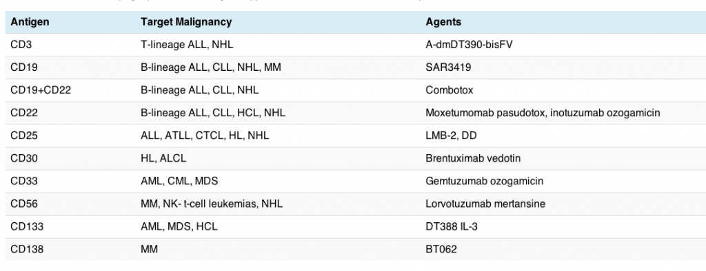
Antibodies that target two antigens are known as bispecific antibodies. Only one is currently available commercially (catumaxomab, Removab) and binds to CD3 and EpCam, although there are several in late stage development, including blinatumomab (Amgen) in ALL. The latter is interesting because it is part of the new generation of antibodies known as bi-specific T-cell engagers (BiTEs).

A bispecific monoclonal antibody (BsAb) is a manufactured protein that is composed of fragments of two different monoclonal antibodies and consequently binds to two different types of antigen.

Manufacturing a monoclonal antibody, while more complex than an oral tyrosine kinase inhibitor (TKI), is easier than a bispecific antibody. Much of the limitations seen so far with bispecific antibodies have been technological rather than clinical. What the Genentech scientists set out to do is succinctly described by Dr Scheer in the short Soundcloud below:

What are the advantages of bispecific antibodies?

The main advantage of bispecific antibodies is the ability to combine a cytotoxic cell (e.g. CD3) or ADC with a tumour specific protein target (e.g. CD19 or CD20) although a number of different combinations could be considered. In other words, you would get the ability to home in on the specific tumour target together with enhanced cell killing.

This could be a potent combination, except that technology-wise, they are difficult to engineer as Speiss and colleagues noted:

“… bispecific-antibody design and production remain challenging, owing to the need to incorporate two distinct heavy and light chain pairs while maintaining natural nonimmunogenic antibody architecture.”

There are some technological difficulties in engineering bispecific antibodies, though.  Blinatumomab was mentioned as one example by Speiss et al., (2013) because:

“… some bispecific antibody fragments (e.g., the anti-CD19-CD3 single-chain fragment blinatumomab) are expressed as a single polypeptide chain they include potentially immunogenic linkers.”

What was fascinating about the Nature Biotech paper was that they reported on a new process they had developed to manufacture bispecific antibodies:

“We present a bispecific-antibody production strategy that relies on co-culture of two bacterial strains, each expressing a half-antibody.  Using this approach, we produce 28 unique bispecific antibodies.”

One thing I thought was particularly cool about this novel approach is that bacteria are easier to manipulate and having a foreign component in the antibody will potentially mean that the human body’s immune system will hopefully pick it up more easily. Essentially, these new chemical structures could act as a powerful cancer homing device against specifically chosen targets.

The example used in the paper was a new bispecific antibody they engineered from co-cultures of EGFR and MET. Remember that Genentech/Roche already has a TKI against EGFR (erlotinib) on the market and a MET antibody (onartuzumab) in development. Neither of these drugs hit both targets and yet as Speiss and colleagues noted:

“MET and EGFR drive the growth of a marked proportion of non-small cell lung cancer tumors. MET and EGFR are often co-expressed and co-activated, and MET signaling can compensate for loss of EGFR signaling and vice versa.”

Image Courtesy of Roche's gRED unit: Bispecific antibody with two distinct binding arms that inhibits both MET (orange) and EGFR (green). The bispecific antibody, shown here in red and blue, has a natural antibody architecture

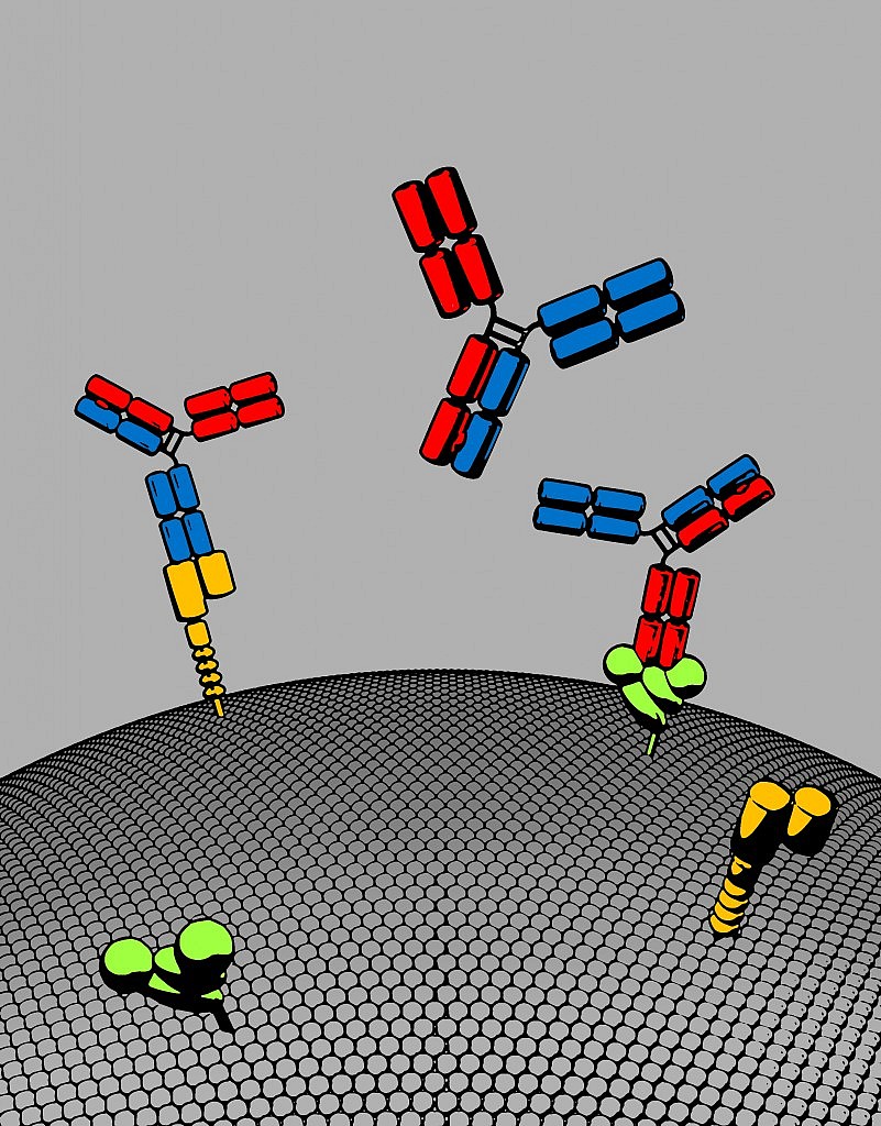
Image Courtesy of Roche’s gRED unit: Bispecific antibody with two distinct binding arms that inhibits both MET (orange) and EGFR (green). The bispecific antibody, shown here in red and blue, has a natural antibody architecture

As Dr Scheer observed, we don’t know yet is where the company will go with this exciting technology, but if the approach shows promising efficacy in future clinical trials, then it’s easy to see how multiple new bispecific antibodies could be easily developed for different tumour types, with far more potency and utility than single targeted therapies alone.

Stop and think about that possibility for a moment.

Transformative science isn’t always about finding a new target, sometimes the breakthrough is in removing the technological limitations to create a much more robust platform with enormous therapeutic potential.  At that point, the biology, targets and imagination become the limitations, not the technology itself.

I have a feeling that this platform is a much more exciting breakthrough than many realise – it’s the sort of approach where you can see, to paraphrase a famous watch company’s ad – some day all antibodies will be made this way.

References:

ResearchBlogging.orgSpiess C, Merchant M, Huang A, Zheng Z, Yang NY, Peng J, Ellerman D, Shatz W, Reilly D, Yansura DG, & Scheer JM (2013). Bispecific antibodies with natural architecture produced by co-culture of bacteria expressing two distinct half-antibodies. Nature biotechnology PMID: 23831709

The Cancer Research journal has an interesting review of ADC and immunotoxin technology and their potential role in hematologic malignancies that is well worth reading (see references below).

Fitzgerald et al., (2011) observe that:

Immunotoxins and ADCs are assembled in a number of different ways. Antibody fragments or whole antibodies are combined with either protein toxins or low-molecular-weight toxic drugs. Linkage options include gene fusions (peptide bonds), disulfide bonds, and thioether bonds.

Design goals dictate that immunotoxins and ADCs remain intact while in systemic circulation but disassemble inside the target cell, releasing the toxic payload.

Uncoupling the toxin or drug from the antibody is accomplished by protease degradation, disulfide bond reduction, or hydrolysis of an acid-labile bond. Toxin or drug attachment to the antibody must not interfere with antigen binding.

I was particularly struck by the number of immunotoxins and ADCs currently in clinical development each focused on different targets. The article highlights a number of agents in the clinic, as of January this year.

A quick overview is provided in the table below:

ADC

The good news is that some of these agents have already been approved in the US, although not all have seen a successful launch. While the current rising star from Seattle Genetics and Millennium, brentuximab vedotin (Adcetris), received fast track approval from the FDA in HL and ALCL earlier this year, gemtuzumab ozogamicin (Mylotarg) was voluntarily withdrawn from the US market by Pfizer last summer.  Part of Mylotarg’s problems lay in the early generation linker technology that release the active substance too early, thereby causing more extensive side effects than would be preferred.  Linker technology has evolved considerably since then, which is good news for patients since active drug can now be targeted more precisely where it is most needed.

As the antibody and linkage technology continues to improve, thereby allowing the active drug to be released in the tumour or on the surface of the cancer cells, I would expect to see more of these agents make it to market successfully with fewer toxicities.

One promising agent in this list, moxetumomab pasudotox (MedImmune), had some promising early data presented at ASH in 2010 in relapsed, refractory hairy cell leukemia (HCL) that is worth checking out.

The regular deadline for this years ASH abstracts has already closed, while the late breaker deadline is this month. I’m hoping we will see some more promising data emerge in December.

References:

ResearchBlogging.orgFitzGerald, D., Wayne, A., Kreitman, R., & Pastan, I. (2011). Treatment of Hematologic Malignancies with Immunotoxins and Antibody-Drug Conjugates Cancer Research, 71 (20), 6300-6309 DOI: 10.1158/0008-5472.CAN-11-1374

The beauty of social media is that sometimes someone shares something monumental before you even pick it up yourself in a journal you’re subscribed to. I love that – it’s a great way to see how others find things with what I call ‘interestingness’.  This morning, John Carroll of Fierce Biotech tweeted something that gave me goosebumps.

What was hot this morning you may all be wondering?

Crossing the blood-brain barrier (BBB)?

Wow, now that is something that really grabs my attention!  Normally, when you try to target drugs to the brain, you find that the endothelium acts as an impenetrable and largely impervious layer that forces you to keep adding more and more drug in an effort to get a small amount through.  This approach obviously increases side effects and the reality is so little drug actually gets through that efficacy is severely limited.

It also explains why we haven’t made much progress with brain related diseases such as Alzheimer’s, Parkinson’s and Glioblastoma (a malignant brain cancer).

We’ve talked a bit about nanotechnology on this blog and it’s companion, Biotech Strategy Blog, as a way to make things small enough to potentially cross the BBB, but I was keen to see what this was all about.  Enthusiastically, I checked out the original article in Science and Translational Medicine myself (see references below).  The editorial commentary associated with it began:

“As impenetrable as the walls of ancient Troy, the tight endothelial cell layer of the blood-brain barrier (BBB) allows only a few select molecules to enter the brain. Unfortunately, this highly effective fortress blocks passage of therapeutic antibodies, limiting their usefulness for treating diseases of the brain and central nervous system.”

Oof, a tad dramatic perhaps, but what did the Genentech scientists do that was different?

Well, the research team were looking at using a BACE-1 antibody to block the enzyme involved in amyloid production, but the BBB prevented little drug from getting through, despite higher doses.

The engineers then developed a new antibody to take advantage of the fact that cells need iron by creating an antibody with two arms:

  • One arm held anti-BACE1 drug
  • The other arm hosted a receptor called transferrin that carries iron to brain cells, providing a ferry across the barrier

In other words, they made use of the body’s natural transport system in much the same way the Trojan horse carried men.  Perhaps the analogy wasn’t so dramatic after all…

This novel approach allowed the scientists to use lower concentrations of the new drug to get the active therapy through.  This should limit side effects and hopefully, increase efficacy.  According to the authors:

“BACE1 can be targeted in a highly selective manner through passive immunization with anti-BACE1, providing a potential approach for treating Alzheimer’s disease. Nevertheless, therapeutic success with anti-BACE1 will depend on improving antibody uptake into the brain.”

Note my emphasis… I can just see the anti-immunisation crowd up in arms before we even get started on this.

Implications of this research

The obvious potential benefits from this novel approach are in Alzheimer’s Disease research, although I caution that we still need to see results from human trials.

However, what piques my interest is how the technological approach of the iron receptor could be used for other brain diseases such as Parkinson’s Disease and brain tumours such as glioblastoma (GBM), which is a nasty malignant cancer limited by how much drug we can get inside the tumour cells. It crossed my mind that it might not take long for Genentech scientists and engineers to use the iron transport concept to bolt together a new monoclonal antibody combining the transferrin receptor and bevacizumab (Avastin), which is already approved as a treatment for GBM. Who knows how that might pan out, but the idea is very appealing indeed.

It is the development and spreading of novel and creative ideas like these that really excites me as a scientist and reminds me what I loved about it as kid.

Crazy Deranged Fools

This idea is so creative, so simple, so brilliant that I’m giving the Genentech scientists my Crazy Deranged Fool (CDF) award of the month for having the temerity to try something so boldly different – check out Hugh’s cartoon at Gaping Void to find out what that is!

References:

ResearchBlogging.orgAtwal, J., Chen, Y., Chiu, C., Mortensen, D., Meilandt, W., Liu, Y., Heise, C., Hoyte, K., Luk, W., Lu, Y., Peng, K., Wu, P., Rouge, L., Zhang, Y., Lazarus, R., Scearce-Levie, K., Wang, W., Wu, Y., Tessier-Lavigne, M., & Watts, R. (2011). A Therapeutic Antibody Targeting BACE1 Inhibits Amyloid-  Production in Vivo Science Translational Medicine, 3 (84), 84-84 DOI: 10.1126/scitranslmed.3002254

1 Comment

The news yesterday from Amgen that panitumumab (Vectibix) failed in Head and Neck cancer got me thinking.  Why did it fail where cetuximab (Erbitux) succeeded? They're both monoclonal antibodies to EGFR, so that makes it rather interesting.

Many of you will remember that bevacizumab (Avastin) and Erbitux were both approved within a month of each other for colorectal cancer, both monoclonals, to VEGF and EGFR respectively.  That was after a long string of failures for anti-angiogenesis compounds.  Subsequently, vatalanib (PTK787), a small molecule tyrosine kinase inhibitor (TKI) of VEGF from Novartis failed in the same indication and did not achieve statistical significance in survival.

It gets even more interesting when you consider prostate cancer as another example:

  1. Abbott's atrasentan got villified at it's ODAC meeting and never received approval.  In a few months, however, we will hear what happens in the phase III trial for another endothelin-A inhibitor, zibotentan (AstraZeneca).  The phase II data showed a difference in OS, but not PFS, so who knows what will happen with the phase III study?
  2. Avastin also failed to achieve a survival advantage in prostate cancer, but does that mean that other VEGF inhibitors, such as sanofi-aventis/Regeneron's aflibercept (VEGF-Trap), will fall the same fate?  We don't know yet, but as far as I know, that one's still alive and kicking.

I find it fascinating trying to work out why some drugs work and some don't even in the same class in the same indication.  It could be all sorts of reasons:

  • Dosing
  • Scheduling
  • Patient population
  • Study design
  • Drug combinations
  • Compound structure
  • Availability of suitable biomarkers
  • Indications

Or one of many many other things or combination of reasons.

And there there is the related issue of 'pure' inhibitors (single target such as VEGF or BCR-ABL) versus multi-kinases or monoclonals (more than one target such as VEGF, EGFR, PDGF or FGFR, for example).  Does it make a difference in efficacy and tolerability, will they affect outcomes differently?

Hopefully, we can learn from the failures to date for the future, allowing us to design better drugs and trials that have a more positive impact on outcomes.

Curiousity is killing this cat… R&D is such a crapshoot sometimes.


While reading my pile of mail on Friday, I realised that an interesting paper on Hodgkins Lymphoma (HL) appeared in the current edition of the New England Journal of Medicine (full reference below).

The basics of the paper are that despite advances in HL, including curative radiation in the early stages if the disease, one third of patients with advanced disease and about 15% of those with early disease have a relapse after treatment. 20% of people still die from the disease.  

The question is why?  

Well, unfortunately, current prognostic models have not been shown to be very accurate and so far, no biomarker has been found to be particularly useful.

The authors set out to use gene-expression profiling obtained from people with HL during diagnostic lymph node biopsy to determine which signatures were correlated with treatment.  They confirmed their findings with an independent cohort of 166 patients using standard immunohistochemical analysis.

What they found was fascinating:

  • A gene signature of of tumour associated macrophages was associated with primary treatment failure.
  • An increased number of CD68 macrophages correlated with likelihood of relapse after autologous stem cell transplantation.
  • The adverse prognostic factor (macrophages) outperformed the current International Prognostic Score for disease-specific survival.
  • The absence of an elevated number of CD68+ cells in patients with limited stage disease defined a sub-group of patients with long-term disease specific survival of 100% with the current therapies.

When I was at school, we learned that macrophages were associated with an immune response to invasion.  According to the NEJM Editorial, by DeVita and Costa, this may not be the case:

"Most of the evidence, however, now links the presence of tumor-associated macrophages with a poor prognosis."

In short, the data shows that increased number of tumour associated macrophages was strongly associated with shortened survival in HL and provides a biomarker for prognosis and risk stratification.  What does this all mean though, for clinical practice?

DeVita and Costa noted that:

"If at the time of diagnosis we could identify patients who are destined to have a poor response to treatment, most patients could be spared a combination of therapies or radiotherapy with its attendant long-term toxic effects."

This is an important observation alone.


For the future, though, the data suggests some new directions that clinical research could go in, such as an anti-CD68 monoclonal antibody perhaps.  There are some that have been identified for rheumatoid arthritis (RA) as the Kunisch paper shows below, but I don't think any are currently in commercial development at the moment.

For the moment, though, I'm left wondering more than there are answers.  

Why do people with macrophages do worse, what is the mechanism for this?  How can we best target the macrophages or the CD68 cells?  If people are screened and are found to have a poorer prognosis and are spared the exposure to chemotherapy or radiation as DeVita and Costa suggest, how should they be treated instead?

Perhaps more research will be galvanised by Steidl et al's findings.  Time will tell.


ResearchBlogging.org
Steidl C, Lee T, Shah SP, Farinha P, Han G, Nayar T, Delaney A, Jones SJ, Iqbal J, Weisenburger DD, Bast MA, Rosenwald A, Muller-Hermelink HK, Rimsza LM, Campo E, Delabie J, Braziel RM, Cook JR, Tubbs RR, Jaffe ES, Lenz G, Connors JM, Staudt LM, Chan WC, & Gascoyne RD (2010). Tumor-associated macrophages and survival in classic Hodgkin's lymphoma. The New England journal of medicine, 362 (10), 875-85 PMID: 20220182


DeVita, V., & Costa, J. (2010). Toward a Personalized Treatment of Hodgkin's Disease New England Journal of Medicine, 362 (10), 942-943 DOI: 10.1056/NEJMe0912481

Kunisch, E. (2004). Macrophage specificity of three anti-CD68 monoclonal antibodies (KP1, EBM11, and PGM1) widely used for immunohistochemistry and flow cytometry Annals of the Rheumatic Diseases, 63 (7), 774-784 DOI: 10.1136/ard.2003.013029

error: Content is protected !!