Pharma Strategy Blog

Commentary on Pharma & Biotech Oncology / Hematology New Product Development

Posts from the ‘Strategy’ category

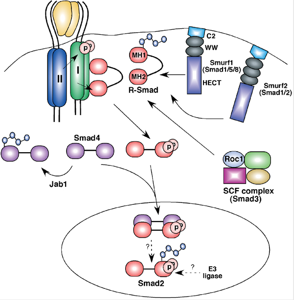
“Ubiquitin-dependent mechanisms have emerged as essential regulatory elements controlling cellular levels of Smads and TGFβ-dependent biological outputs such as epithelial–mesenchymal transition (EMT).

In this study, we identify a HECT E3 ubiquitin ligase known as WWP2 (Full-length WWP2-FL), together with two WWP2 isoforms (N-terminal, WWP2-N; C-terminal WWP2-C), as novel Smad-binding partners. We show that WWP2-FL interacts exclusively with Smad2, Smad3 and Smad7 in the TGFβ pathway.

Interestingly, the WWP2-N isoform interacts with Smad2 and Smad3, whereas WWP2-C interacts only with Smad7. In addition, WWP2-FL and WWP2-C have a preference for Smad7 based on protein turnover and ubiquitination studies. Unexpectedly, we also find that WWP2-N, which lacks the HECT ubiquitin ligase domain, can also interact with WWP2-FL in a TGFβ-regulated manner and activate endogenous WWP2 ubiquitin ligase activity causing degradation of unstimulated Smad2 and Smad3.

Consistent with our protein interaction data, overexpression and knockdown approaches reveal that WWP2 isoforms differentially modulate TGFβ-dependent transcription and EMT.

Finally, we show that selective disruption of WWP2 interactions with inhibitory Smad7 can stabilise Smad7 protein levels and prevent TGFβ-induced EMT.

Collectively, our data suggest that WWP2-N can stimulate WWP2-FL leading to increased activity against unstimulated Smad2 and Smad3, and that Smad7 is a preferred substrate for WWP2-FL and WWP2-C following prolonged TGFβ stimulation.

Significantly, this is the first report of an interdependent biological role for distinct HECT E3 ubiquitin ligase isoforms, and highlights an entirely novel regulatory paradigm that selectively limits the level of inhibitory and activating Smads.”

Source: Oncogene

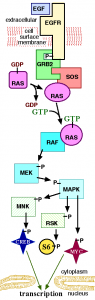
That was an abstract I was browsing over coffee in my oncology RSS feeds and while it was a bit heavy for early in the day, I was intrigued because Smads have been cropping up in GI sessions at meetings over the last six months or so.  Smads are signal transducers for members of the transforming growth factor-beta (TGF-beta) superfamily, so they occupy a key role in transcription of proteins:

The biology of TGF-beta and Smads

In addition, I’ve included a link to an open access article on the biology of Smads in the references below.

Essentially, the translational research from Soond and Chantry (2011) is suggesting that blocking the WWP2 gene could prevent metastasis, ie cancers from spreading to other organs of the body.  Many of you will remember the post on Norton and Massague’s cancer cell seeding theory and this new finding could well have implications for that research too.

Overall, the latest findings mean that if we have a valid target, we can design a drug to target the rogue gene sending signals.

Of course, these are still very early days yet, but it will be interesting to see if the basic science can be translated into R&D and eventually, a real clinical impact in the long run.

For those of you wanting a simpler version of the abstract, BBC Health did a nice job of putting the research into plain English.  Do check out their short report with pretty pictures here.

References:

ResearchBlogging.orgSoond, S., & Chantry, A. (2011). Selective targeting of activating and inhibitory Smads by distinct WWP2 ubiquitin ligase isoforms differentially modulates TGFβ signalling and EMT Oncogene DOI: 10.1038/onc.2010.617

Attisano, L., & Tuen Lee-Hoeflich, S. (2001). The Smads Genome Biology, 2 (8) DOI: 10.1186/gb-2001-2-8-reviews3010

Izzi, L., & Attisano, L. (2004). Regulation of the TGFβ signalling pathway by ubiquitin-mediated degradation Oncogene, 23 (11), 2071-2078 DOI: 10.1038/sj.onc.1207412

On Thursday this week I’m off to the GI Carcinogenesis meeting hosted by MD Anderson Cancer Center, you can find out more about the event here.

It’s a brand new meeting for me, but according to the program:

“The ISGC is comprised of basic, translational and clinical scientists.  This conference will encourage and develop research and communication in the areas of gastroenterological biology and oncology in both basic and clinical aspects through joint meetings with international and national gastroenterologists.”

I’m particularly looking forward to hearing what Lee Ellis has to say on cancer stem cells and the microenvironment, as well as Emanuel Petricoin on molecular profiling in GI cancers.  There are a whole host of other really interesting talks too, as you can see from the program agenda.

When I first looked at the faculty, my initial reaction was, “Oh my!” It’s a quite serious line-up of some of the top GI cancer researchers and certainly not easy to get them all in the same place together, so it will be fun to chat with them in the poster sessions and get their perspective on the latest happenings in this field.

The meeting runs through Saturday, so I’ll try and post a daily synopsis, as time permits.

If I hadn’t been following Dr Raymond DuBois, the MD Anderson Provost and Co-Chair of the meeting on Twitter, I would have missed this altogether – the power of social media in spreading and communicating awareness of these special events is very much here to stay.

If you have any burning questions in this area, please do add them in the comments below and I will do my best to ferret out some answers.

1 Comment

One of my favourite journals, Cancer Research, has a new paper available via open access (i.e. free to the public, thank you AACR), which you can obtain from the link in the Reference section below.

It caught my attention because there was a fascinating symposium on angiogenesis at ESMO this summer with some heavyweight debates from Robert Kerbel (accelerated metastasis) and Lee Ellis (normalisation of tumour vessels) taking different viewpoints on the pros and cons of VEGF inhibition.  I took a few photos of the slides for private study and reflection, as they were going too fast for me to keep up with the key points with unreadable chicken scratch notes, but sadly my iPhone went missing in the exhibit hall less than an hour afterwards before I could download the photos :(.  That said, both sides argued with very compelling data for their perspective that I’m not sure which way I roll on the issue.

In this latest paper, di Tomaso et al., from Boston discuss the concept of recurrent glioblastomas and the tendency to relapse after VEGF therapy.  They noted that there are two current theories for how this might happen:

  1. Switch to VEGF-independent angiogenic pathways
  2. Vessel co-option

They therefore decided to investigate these mechanisms in patients with relapsed glioblastoma using a pan VEGF inhibitor, cediranib.  Now, it should be noted that cediranib (Recentin) is not yet approved and is a small molecule inhibitor, whereas another VEGF inhibitor, bevacizumab (Avastin), is a monoclonal antibody approved for relapsed GBM, so I’m sure why they didn’t use that instead.  It does make extrapolation of the findings a little more tricky though, as you cannot always assume a class effect.

Here are the key findings:

  • Endothelial proliferation and glomeruloid vessels were decreased
  • Vessel diameters and perimeters were reduced to levels comparable to the unaffected contralateral brain hemisphere
  • Tumour endothelial cells expressed molecular markers specific to the blood–brain barrier, indicative of a lack of revascularization despite the discontinuation of therapy
  • Cellular density in the central area of the tumour was lower than in control cases and gradually decreased toward the infiltrating edge, indicative of a change in growth pattern of relapsed GBM after cediranib treatment
  • Cediranib-treated GBMs showed high levels of PDGF-C (platelet-derived growth factor C) and c-Met expression and infiltration by myeloid cells, which may potentially contribute to resistance to anti-VEGF therapy

The authors therefore concluded that:

“rGBMs switch their growth pattern after anti-VEGF therapy—characterized by lower tumor cellularity in the central area, decreased pseudopalisading necrosis, and blood vessels with normal molecular expression and morphology—without a second wave of angiogenesis.”

Commentary:

What intrigued me in particular was not the lack of rebound vascularisation effect but the myeloid component.  Many of you will remember the AACR meeting last September on Molecular Diagnostics in Cancer Therapeutics, where AVEO presented data on their VEGF inhibitor in development and found that the myeloid component acted as a useful biomarker of response for tivozanib in renal cell cancer. You can read more about that here if you missed it.

This raises several interesting questions for me:

  1. Is the myeloid marker that AVEO found with tivozanib actually more useful and applicable to VEGF therapies in general?
  2. Does the myeloid component indicate acute inflammation, as we have seen with respiratory and other diseases?
  3. If PDGF and MET expression rise as resistance sets in, does that suggest logical combination therapies for the treatment of GBM?
  4. How can we better overcome the blood brain barrier, which is a physical impediment to improving outcomes.

Time will tell but clearly the research in relapsed GBM has a-ways to go before we figure out how best to approach it yet.

References:

ResearchBlogging.org di Tomaso, E., Snuderl, M., Kamoun, W., Duda, D., Auluck, P., Fazlollahi, L., Andronesi, O., Frosch, M., Wen, P., Plotkin, S., Hedley-Whyte, E., Sorensen, A., Batchelor, T., & Jain, R. (2011). Glioblastoma Recurrence after Cediranib Therapy in Patients: Lack of “Rebound” Revascularization as Mode of Escape Cancer Research, 71 (1), 19-28 DOI: 10.1158/0008-5472.CAN-10-2602

Earlier this year we discussed some interesting papers on circulating tumour cells (CTC’s) in prostate cancer and how they are becoming a potentially useful surrogate marker in clinical trials for other cancers including lung cancer.

I was therefore intriqued to see another paper looking at the role of CTC’s in colorectal cancer (CRC) – see link below in the reference section at end of the article:

“Circulating tumour cells early predict progression-free and overall survival in advanced colorectal cancer patients treated with chemotherapy and targeted agents”

One of the challenges with some targeted therapies such as bevacizumab (Avastin) now routinely used for treatment of metastatic colorectal cancer is that while they improve overall survival, not all patients will actually respond to treatment.

While several studies have shown that the KRAS mutation status predicts response to EGFR therapy such as cetuximab (Erbitux) and panitumumab (Vectibix) in colorectal cancer, there is no known equivalent biomarker for determining who is most likely to respond to VEGF therapy with bevacizumab.

This situation creates a dilemma for the medical oncologist, because other than excluding those patients most at risk from the side effects (patients with cardiovascular disease, hypertension etc), perhaps 1 in five of the advanced CRC patients, there is no way to determine which of the remaining 4/5 people will respond, thus potentially exposing all to the not insignificant systemic effects of the drug with no idea who might be an ideal candidate.

Previously, research from Cohen et al., has shown in advanced CRC that the CTC at baseline and during treatment were prognostic for OS and PFS (see references below). These studies included a broad heterogeneous population of untreated and pre-treated patients who received different schedules of treatment, making it difficult to draw specific conclusions.  That said, the research demonstrated that the technique is useful and can be measured from blood samples while not requiring invasive biopsies.

In this article, the researchers decided to see whether CRC’s would be a useful tool for predicting the responders from non-responders better in a more homogenous population.

What did the results show?

Perhaps the most interesting quote in this journal article was the finding that:

“The combined analysis of CTC and CT imaging provided a more accurate outcome assessment than either modality alone.”

Clearly, RECIST measurements are not going to go away, but combining the data with newer biomarker analysis that reflects the underlying biology may well be a good compromise.

What does this all mean?

Historically, physicians have used pathologic measures of measuring tumour response using RECIST criteria, but the problem with this approach is that tumour shrinkage alone does not always translate into an improvement in meaningful outcome for the patient. Sometimes patients can have no shrinkage and stable disease but still a reasonable and functional quality of life, as happens with some soft tissue sarcomas, for example.

New surrogate markers of survival are therefore needed that also tell us something about about the risk of recurrence.   This new paper provides additional evidence that:

“The CTC count before and during treatment independently predicts PFS and OS in ACC patients treated with chemotherapy plus targeted agents and provides additional information to CT imaging.”

They based this conclusion on the results that demonstrated:

“The sensitivity and specificity of high CTC at baseline for the prediction of progressive disease on CT imaging were 16.7% and 70.1%, respectively, and of high CTC at 1–2 weeks after the start of treatment 20.0% and 95.1%, respectively.”

They also went on to note that:

“We demonstrate that CTC counts identify a small group of patients with unfavourable outcome early during treatment. However, whether a change in treatment on the basis of CTC count will result in a better survival for this group is yet unknown, and this issue should be addressed in a prospective trial.  In such a design, it will also be worthwhile to investigate the cost-effectiveness of CTC testing.”

Unfortunately, although a reasonably large number patients were evaluated (n=467) in this study, half were treated with capecitabine, oxaliplatin, and bevacizumab and half received the same regimen plus weekly cetuximab, but the results were oddly described in the aggregate.  We therefore have no idea whether CTC’s were more useful in the bevacizumab only arm, or when cetuximab was added.  We do know that in the patients who did poorly, although their specific treatment is not described, they did have high CTC levels and poor disease control.

CTC’s are something we will likely hear a lot more about in research going forward from a biomarker perspective though, as researchers begin to incorporate their measurement into the design of more clinical trials.

References:

ResearchBlogging.orgTol, J., Koopman, M., Miller, M., Tibbe, A., Cats, A., Creemers, G., Vos, A., Nagtegaal, I., Terstappen, L., & Punt, C. (2009). Circulating tumour cells early predict progression-free and overall survival in advanced colorectal cancer patients treated with chemotherapy and targeted agents Annals of Oncology, 21 (5), 1006-1012 DOI: 10.1093/annonc/mdp463

Cohen, S., Punt, C., Iannotti, N., Saidman, B., Sabbath, K., Gabrail, N., Picus, J., Morse, M., Mitchell, E., Miller, M., Doyle, G., Tissing, H., Terstappen, L., & Meropol, N. (2009). Prognostic significance of circulating tumor cells in patients with metastatic colorectal cancer Annals of Oncology, 20 (7), 1223-1229 DOI: 10.1093/annonc/mdn786

Cohen, S., Punt, C., Iannotti, N., Saidman, B., Sabbath, K., Gabrail, N., Picus, J., Morse, M., Mitchell, E., Miller, M., Doyle, G., Tissing, H., Terstappen, L., & Meropol, N. (2008). Relationship of Circulating Tumor Cells to Tumor Response, Progression-Free Survival, and Overall Survival in Patients With Metastatic Colorectal Cancer Journal of Clinical Oncology, 26 (19), 3213-3221 DOI: 10.1200/JCO.2007.15.8923

4 Comments

I’m on a lung cancer and systems biology roll at the moment, although partly that’s just how the interesting data rolls in the literature.

Here’s some new food for thought.  A group of respectable scientists published some fascinating data in PLOS Medicine (free article see reference below) entitled, “Nuclear Receptor Expression Defines a Set of Prognostic Biomarkers for Lung Cancer.”

Using PCR, they evaluated NR expression patterns associated with good and poor outcomes in patients with non-small cell lung cancer (NSCLC) and then validated the findings in lung adenocarcinomas (n=550) and squamous cell carcinoma (n=130) samples in three different analyses by comparing normal and lung cancer cells.  Two important factors emerged from the analysis:

“The prognostic signature in tumors could be distilled to expression of two nuclear receptors, short heterodimer partner (SHP) and progesterone receptor, as single gene predictors of NSCLC patient survival time, including for patients with stage I disease.”

The SHP protein was the better predictor of outcomes in patients with stage I disease; those with strong SHP expression had better overall survival rates of approx. 70% at 100 months compared with 45% among people with low SHP expression.  The survival curves in the paper were quite dramatic – check them out.  Interestingly, the same signatures were also predictive of recurrence based on normal tissue samples from the patients with NSCLC.  Progesterone receptor expression was, however, a much weaker predictor of any outcome based on this analysis.

Essentially, this means the study demonstrated:

“NR expression is strongly associated with clinical outcomes for patients with lung cancer, and this expression profile provides a unique prognostic signature for lung cancer patient survival time, particularly for those with early stage disease.”

What are nuclear receptors, you may be wondering?

“The NR superfamily contains 48 transcription factors (proteins that control the expression of other genes) that respond to several hormones and to diet-derived fats.  NRs control many biological processes and are targets for several successful drugs, including some used to treat cancer.”

Still, it’s not something that immediately springs to mind as a possible or logical prognostic biomarker.

That said, out of the 48 transcription factors, two were found to be related to poorer patient outcomes.  They were NGFIB3, a receptor associated with nerve growth factor, and NR3C2, a mineralocorticoid receptor protein:

“This study highlights the potential use of Nuclear Receptors (NRs) as a rational set of therapeutically tractable genes as theragnostic biomarkers, and specifically identifies short heterodimer partner and progesterone receptor in tumors, and NGFIB3 and MR in non-neoplastic lung epithelium, for future detailed translational study in lung cancer.”

Going forward, we still need to see more research to find out whether these particular NRs or others were involved with tumour development and growth.  If  they do, then NR’s may potentially offer new therapeutic targets for future research and development.

References:

ResearchBlogging.org Jeong, Y., Xie, Y., Xiao, G., Behrens, C., Girard, L., Wistuba, I., Minna, J., & Mangelsdorf, D. (2010). Nuclear Receptor Expression Defines a Set of Prognostic Biomarkers for Lung Cancer PLoS Medicine, 7 (12) DOI: 10.1371/journal.pmed.1000378


At the annual NY Chemotherapy Foundation symposium the other week, one of the highlights for me was listening to Larry Norton (MSKCC) give a 30 minute keynote entitled:

“Cancer Cell Seeding: a hypothesis with therapeutic implications”

He talked at length about what he described as some of the breast cancer mysteries, namely the puzzles associated with:

  1. Phenotypic consistency (hyperplasia, anaplasia, rapid growth, angiogenesis, large tumour size, invasion, metastases and latency)
  2. Disorderly order
  3. Unclear clear margins

Several main points were then raised from this quick synopsis:

  • The phenotypic elements are so inextricably linked that they must be related, but how?
  • Sentinel node mapping proves that progression is orderly. Yet it isn’t! How?
  • RT must kill cells that are beyond clear margins.  How do they get there? What is their relationship to distant recurrence?
  • That RNA expression is prognostic and predictive challenges the notion of tumour-initiating cells being rare.

These issues or assumptions may be true or not true so the question then becomes how are we going to solve them?

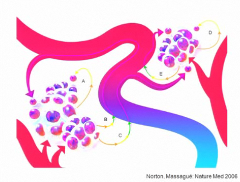
Norton noted that one way these concepts may be related is that the tumour is essentially metastasing to itself and showed a nice schematic from 2006 that he and Massague used in their Nature Medicine paper.  Tumours thus grow by metastasis back to itself and will promote increased growth and bigger tumour size:

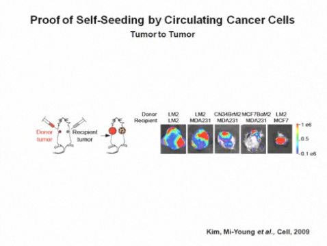
However, it took until late 2009 to research and publish proof that this idea is true (see reference below for the paper):

The other concept that caught my attention is the idea of a ‘toxic sponge’ that followed on from this as Norton was putting the story back together in terms of breast cancer.  In short, the primary tumour and region act as a sponge for circulating tumour cells.  When the sponge is removed or sterilised, circulating tumour cells seek distant sites since they cannot metastasise back to the primary tumour anymore.  Seeding the new sanctuary sites may also create reservoirs for feeding future distant metastases.  Research is now underway to determine whether RT has accelerates or potentiates the metastasis process in the long run.

All in all, a very interesting and well put together talk.

ResearchBlogging.org Norton L, & Massagué J (2006).  Is cancer a disease of self-seeding? Nature medicine, 12 (8), 875-8 PMID: 16892025

Kim, M., Oskarsson, T., Acharyya, S., Nguyen, D., Zhang, X., Norton, L., & Massagué, J. (2009).  Tumor Self-Seeding by Circulating Cancer Cells Cell, 139 (7), 1315-1326 DOI: 10.1016/j.cell.2009.11.025

3 Comments

Metastatic melanoma is quite a hot topic right now with a rich pipeline of products in development after a decade of little or no progress.  Of course, it is a bit like three London buses coming along at once after an hour long wait in the winter weather, but better late than never.

Many of you will remember the recent data from ipilimumab (BMS), an immunotherapy that showed increased survival, albeit with some severe adverse events, from the phase III trial in newly diagnosed metastatic melanoma presented at ASCO in the plenary session earlier this year, followed by a publication in the NEJM.  The FDA filing was subsequently submitted on the basis of the positive data.

Yesterday, BMS announced that the FDA have moved the PDUFA date back 3 months from Dec 25th to March 26th, 2011.  A precise reason for the delay wasn’t given , but the company did say:

“In response to an FDA request, Bristol-Myers Squibb submitted further analysis of data pertaining to the current application for pre-treated advanced melanoma and the agency considers this to be a major amendment to the drug’s BLA.”

I’m not going to speculate on the reasons for the extra review time or what the new data was, but it is an interesting and unexpected development.

Meanwhile, there’s also been a lot of buzz around targeted BRAF inhibition in melanoma lately, specifically around the initial stunning results seen with PLX4032 (Plexxikon & Roche).  So far, it seems that responses of around 6-12 months, with a median of around 8 months are possible with an kinase inhibitor that specifically targets the V600E mutation associated with BRAF, although there two problems:

  1. The responses are not durable as resistance (eg associated with MEK or AKT amplification) sets in.
  2. Inhibiting CRAF as well as BRAF appears to lead to an unwanted excess proliferation of squamous cells, which is reversible on withdrawal of treatment.

In the first case, a couple of recent papers have looked at mechanisms of resistance around BRAF inhibition that give us some clues of where to go next.

Gopal et al., (2010) decided to see what happened with AZD6244 or selumetinib (Array and AstraZeneca), a MEK and MAP/ERK inhibitor, and whether it would have any impact in mitigating BRAF resistance, given the potential close interaction within the RAS-RAF-MAPK pathway and downstream events that could be impacted through cross-talk and feedback loops:

“We analyzed a panel of Braf mutant human cutaneous melanoma cell lines for their sensitivity to growth and survival inhibition by AZD6244. We compared these effects with the baseline activation status of signaling pathways in the cells, and with AZD6244 treatment–induced changes in signaling networks.

These studies have identified the phosphoinositide 3-kinase (PI3K)-AKT pathway as a critical regulator of the efficacy of AZD6244 in Braf-mutant melanomas, including in cells without baseline activation of the pathway.”

In order to determine possible mechanisms of resistance in the cell lines, they compared the effects of AZD6244 treatment on their signaling pathways with effects in sensitive cell lines and found:

“Although all four of these Braf-mutant cell lines showed similar degree and duration of MAPK inhibition and several other proteins, the resistant cell lines increased their P-AKT levels following exposure to AZD6244, which was not observed in the sensitive cell lines.”

They went on to note:

“The functional significance of AKT activation is supported by the fact that inhibition of AKT activity, either by AKT knockdown or concurrent treatment with the mTORC1/2 inhibitor AZD8055, resulted in synergistic cell killing in the resistant cell lines.”

AstraZeneca and Merck have an ongoing partnership with their MEK (AZD6244) and AKT (MK-2206) kinase inhibitors, so combining them in a clinical trial to try and reduce resistance via feedback loops here would be an interesting approach worth trying.  Such a combination trial is currently recruiting in advanced solid tumours, not melanoma per se.  It is, however, a classic catch-all phase I study to see what kinds of cancers might respond and determine the MTD, but I would be very interested to see the data from patients with metastatic melanoma if they are enrolled.

Now, it has been shown in breast cancer cell lines showed that MEK inhibition resulted in cross-activation of the EGFR tyrosine growth factor receptor, but EGFR has not been shown to be relevant in melanoma, so Gopal et al., considered what other receptors might be responsible for mediating the effects.   In the discussion, an interesting snippet caught my eye:

“AZD6244 treatment induced a slight increase of IGF-I secretion by the cells, and knockdown of IGF-I also blocked P-AKT induction by AZD6244.  Supporting a specific role for the pathway in cell survival, recombinant IGF-I treatment blocked AZD6244-induced cell death, but not growth arrest, in the sensitive WM35.”

This might also suggest another useful combination approach to consider in clinical trials.

Previously, it has been shown that targeting BRAF can not only inhibit the important driver in melanoma, the V600E mutation, but it can also stimulate cellular signaling through the MEK-ERK pathway by activating the related family member C-RAF. This may explain the squamous cell proliferation seen in some patients with PLX4032. The more ideal BRAF inhibitor would therefore specifically target BRAF V600E, without activating CRAF at the same time.

Related to the subject of malignant melanoma, Kamata et al., (2010) just published a paper that looked at the relationship between BRAF and CRAF in the disease.  Previously it has been shown that D594A BRAF lacks kinase activity, but can induce the related gene product CRAF in addition to the mitogen-activated protein/extracellular signal-regulated kinase (ERK) kinase (MEK)/ERK pathway.  What they found was really interesting.  In a nutshell:

“We show that the aneuploid phenotype is dependent on Craf. Treatment with the MEK inhibitor U0126 did not attenuate the emergence of aneuploidy but prevented the growth of aneuploid cells.  These results provide a previously unidentified link between Craf and chromosomal stability, with important implications for our understanding of the development of cancers with driver mutations that hyperactivate Craf.”

Aneuploidy is an abnormal number of chromosomes and can lead to genetic instability, a key cancer hallmark. It’s an important concept here because Kamata et al., have offered a different reason for the CRAF proliferation observed with some BRAF inhibitors:

“Impaired activity BRAF mutants are frequently coincident with oncogenic RAS mutations in human cancers (26) and in these, albeit rare, cancers, we may expect the hyper-activated CRAF induced by the combination of both oncogenes to enhance the aneuploidy response compared with mutation of either oncogene alone.  Such a situation is likely to be highly detrimental to the individual and, indeed, this mechanism may well account for the highly aggressive melanomas we observed following the combined expression of D594A Braf and G12D Kras in melanocytes.”

All in all, this is a very complex yet fascinating area of research and for those of you interested in this field, I would highly recommend reading the latest papers.

Photo Credit: Wikipedia

References:

ResearchBlogging.org Boni, A., Cogdill, A., Dang, P., Udayakumar, D., Njauw, C., Sloss, C., Ferrone, C., Flaherty, K., Lawrence, D., Fisher, D., Tsao, H., & Wargo, J. (2010). Selective BRAFV600E Inhibition Enhances T-Cell Recognition of Melanoma without Affecting Lymphocyte Function Cancer Research, 70 (13), 5213-5219 DOI: 10.1158/0008-5472.CAN-10-0118


Garnett MJ, Rana S, Paterson H, Barford D, & Marais R (2005). Wild-type and mutant B-RAF activate C-RAF through distinct mechanisms involving heterodimerization. Molecular cell, 20 (6), 963-9 PMID: 16364920

Gopal, Y., Deng, W., Woodman, S., Komurov, K., Ram, P., Smith, P., & Davies, M. (2010). Basal and Treatment-Induced Activation of AKT Mediates Resistance to Cell Death by AZD6244 (ARRY-142886) in Braf-Mutant Human Cutaneous Melanoma Cells Cancer Research, 70 (21), 8736-8747 DOI: 10.1158/0008-5472.CAN-10-0902

Kamata, T., Hussain, J., Giblett, S., Hayward, R., Marais, R., & Pritchard, C. (2010). BRAF Inactivation Drives Aneuploidy by Deregulating CRAF Cancer Research, 70 (21), 8475-8486 DOI: 10.1158/0008-5472.CAN-10-0603

4 Comments

Here’s a nice paper that I’ve been reading, written by Tim Harris and Frank McCormick on cancer biology.  Lately, we’ve all seen how advances in DNA sequencing and genome-wide association studies (GWAS) are driving the discovery of the germline and somatic mutations that are present in different cancers.  This article sets out to review:

“The most important molecular changes in different cancers from the perspective of what should be analyzed on a routine basis in the clinic.”

Essentially, this is an overview of where we are in both hematologic and solid tumours and looks at the molecular subsets that are emerging, hopefully as targets for therapeutic intervention.  I’m not going to repeat their excellent article, but if you are really interested in this field, I highly recommend reading it.  A link is provided below after the post to the actual paper.

What struck me most though, was not the nice summary of what we know about the biology of cancer, but their vision of the future in cancer medicine:

“The year is 2020. I wake up and feel the lump under my arm that has been bothering me for several weeks. I decide to make an appointment with the doctor to find the cause, especially since my personal genetic analysis has highlighted alleles that are associated with an increased risk of cancer.

The doctor’s receptionist views my electronic health record online before I am buzzed in to see the doctor.  Once I have explained the problem, a biopsy from the offending lymph-node is taken, the tissue is flash-frozen using the nitrogen quick-freeze system, and then delivered to the laboratory downstairs for a rapid molecular work-up.

DNA from the tissue is sequenced to identify any mutations in the 500 most common genes known to be involved in cancer. Tissue sections are analyzed using high-resolution fluorescent optical images.  A blood sample is also taken to check my background genomic DNA sequence, concentrating on alleles known to predispose to lymphoma.  Sequences for the genes encoding drug-metabolizing enzymes and drug-distribution proteins are also obtained.

A proteomic work-up is undertaken to look at protein profiles and post translational modifications.  I also undergo new-generation imaging so the gross pathology of my organs can be viewed in three-dimensions.  Two hours later, I review the results on my handheld computer device. The results have been predigested and presented as a simple digital read-out so that a diagnosis, prognosis and appropriate treatment can be derived.

Fortunately, the overall molecular and cellular pathology of the lymph-node is considered normal.  Apart from my pre-existing heart disease, all other organs appear to be healthy and I am prescribed an anti-inflammatory drug.

I leave the doctor’s office with a sigh of relief that all appears to be well.”

Whoa, that may sound like something from Bones in Star Trek initially, but I suspect it may well not be as far fetched as we imagine.  Why?   Because over the last two years the progress made in systems biology and cancer genome studies have encouraged me greatly.  What was a fledging area of cancer research is now becoming very much to the forefront of new breakthroughs and increased understanding of what is happening at the molecular level and new prognostic and predictive biomarkers are emerging.

Of course, there is still a long way to go in the war on cancer, but I see plenty of signs that much progress is being made.  Where we may well fall down though, is not in the science per se, but rather in our efforts of communication and coordination:

“The stakeholders, which include the payers, health care organizations, pharmaceutical and biotechnology industry, and molecular diagnostics companies, need to be aligned to achieve the most effective partnership.”

We clearly have much to do in this direction.  We have many of the electronic tools available already and yet speed of testing, diagnosis, access to electronic patient records (EHR) and in particular communication, is often snail mail slow.  How many of us receive test results instantly on our PDAs or email? How many have physicians or other service providers who even communicate with their patients by email or other technologies? This needs to change, and for the better.

There are some nice examples of real empowerment emerging from the cancer community, as this post from my friend Jody, a breast cancer survivor, shows in her recent blog post.  It’s a great start and I hope to hear of many more examples like this.

What do you think?   What can be done to improve the delivery of healthcare for people with cancer and how can we foster greater collaborations?

ResearchBlogging.org
Harris, T., & McCormick, F. (2010). The molecular pathology of cancer Nature Reviews Clinical Oncology, 7 (5), 251-265 DOI: 10.1038/nrclinonc.2010.41

8 Comments

While reading the latest New England Journal of Medicine, it was hard not to notice the focus on ALK mutations and crizotinib (Pfizer), with four articles in all on the topic, including a full original article, two brief reports and an editorial to boot.

Historical background

Gradually, we are gradually seeing more specific molecular markers and targets evolve in non-small cell lung cancer (NSCLC), which is a particularly difficult cancer to treat. This is good news because if we have a relevant target, we can treat subsets of patients with specific drugs and increase their chances of better outcomes while sparing those who are unlikely to respond from the systemic effects of a potent drug. Many call this ‘personalised medicine’ but I prefer to think of it as targeted therapy, because we are essentially matching the drug to the patient’s needs using biomarkers.

The first mutation that evolved in lung cancer, epidermal growth factor receptor (EGFR), was shown to be important in a subset of patients who tested positive for the activating mutation and tend to do well on EGFR therapy with drugs such as erlotinib (OSI/Roche/Genentech) or gefitinib (AstraZeneca).

Originally, two groups reported the discovery of the anaplastic lymphoma kinase (ALK) fusion gene in lymphoma in 1994.  Once translocation has occurred between the EML4 and ALK genes it produces a fusion gene, EML4-ALK, which generates aberrant signaling in cancer.

There was, however, much buzz and excitement three years ago when Japanese researchers reported activating mutations or translocations of ALK in NSCLC. Soda et al’s seminal 2007 paper in Nature is well worth reading from a historical perspective (the link below offers a rare free download from the publishers). As far as I can tell from the literature and talking to lung cancer specialists, the ALK mutation occurs in approx 2-7% of lung cancers. This may sound like a small number, but right now we don’t have anything approved specifically for this particular subset of patients.

Since the Soda paper linking the ALK mutation with lung cancer, we have been fortunate to follow the progress of a compound, originally in development as a c-MET inhibitor, crizotinib. Serendipity is a fine thing sometimes, as the agent had weak Met activity, but Pfizer realised that it had potent ALK activity and sensibly switched the development strategy to investigate it’s possible role in targeting ALK in people with lung cancer. This was a smart decision that is now paying off with a raft of promising data coming out this year at the ASCO and ESMO cancer conferences.

The data from ESMO

Data from a phase I trial was reported in Milan last month.  The study consisted of two parts:

  1. A dose-escalation study that enrolled patients with advanced solid tumors (n=37), including NSCLC, colorectal, pancreatic and inflammatory myofibroblastic tumors (IMT), to determine toxicity and the maximum tolerated dose (MTD).
  2. An expansion cohort, where patients with ALK-positive advanced NSCLC (n=113) were prospectively identified and enrolled with a dose of 250mg twice daily (BID), in order to evaluate the safety and activity of crizotinib in this patient population. All patients had received at least one prior chemotherapy regimen.

Let’s focus on part 2 and the lung cancer patients for brevity.  An important point to note is that majority of patients in the analysis had adenocarcinoma histology and were never or former smokers. The progression free survival (PFS) results were particularly impressive.  At the time of the analysis, preliminary median PFS was 9.2 months (95% CI: 7.6, 10.3), with a median follow-up of 8 months.  Nearly half (47.8%) of the patients with NSCLC remain in follow-up for PFS, while 32% of patients experienced disease progression.  Of those who progressed, 42% continued to receive treatment with crizotinib, as they were determined by investigators to be receiving continued clinical benefit.

The side effect profile showed the agent was well tolerated. The most commonly reported adverse events (all grades) included nausea, diarrhea, mild visual disturbances, and vomiting. Grade 3 ALT (alanine aminotransferase) and AST (aspartate aminotransferase) elevations occurred in four patients.  One patient experienced a grade 4 ALT, another had grade 3 pneumonitis and one patient discontinued the study due to treatment-related adverse events.  Neutropenia, lymphopenia and fatigue were also reported at grade 3 severity (n=6). Compared to standard chemotherapy regimens used to treat NSCLC, I think this looks a much more tolerable and acceptable profile so far.

The NEJM data

The NEJM article from Kwak et al., (2010) describes updated results from the part 2 expansion cohort in the phase I trial described above and offers an updated snapshot of the 82 patients reported at ASCO.  The findings (at the data cut-off) were as follows:

  • The main side effect was mild GI symptoms (grade 1 or 2) and grade 3/4 events were similar to those reported at ESMO.
  • The overall response rate was 57%, with a mean of 6.4 months.
  • There was 1 confirmed complete response (CR) and 46 confirmed partial responses (PR). 27 patients (33%) had stable disease.
  • A total of 63 of 82 patients (77%) were continuing to receive crizotinib.
  • The estimated 6 month PFS rate was 72%.

These data illustrate the value in molecular prescreening large numbers of people with NSCLC to determine who should receive the agent.   Interestingly, the ALK mutation was found in approx. 5% of those who underwent screening. It’s unclear from the paper whether any of the patients who were ALK+ also had an EGFR mutation or whether they are mutually exclusive, but if any patients with NSCLC have both, it would interesting to see if a combination with crizotinib and erlotinib would improve survival.

Overall, these results are both very encouraging and promising for a phase I trial.  Certainly, I haven’t been as excited about such early data since the imatinib (Gleevec) phase I data was presented by Brian Druker (OHSU) in 1999.  Crizotinib is certainly something I will be eagerly following as it progresses through R&D and Pfizer should be applauded for their speedy development in clinical trials once the ALK mutation was discovered and published.

The future

Beyond lung cancer, another article in the NEJM described the impact of crizotinib in treating ALK-rearranged inflammatory myofibroblastic tumour (IMT) (see link below).  This is a rare soft tissue sarcoma arising out of mesenchymal cells and characterised by spindle cell proliferation with an inflammatory element.  Butrynski et al., describe a single case report of a patient with a sustained partial response to crizotinib, thereby potentially offering a new therapeutic strategy for treating these patients.

The Japanese researchers have also described mutations that confer resistance to ALK inhibitors.  Choi et al., have now reported the discovery of two secondary mutations (L1196M and C1156Y) within the kinase domain of ELM4-ALK that conferred resistance to treatment in the NEJM (see link below). This is important, as we have learned from the CML experience, because different inhibitors may target different mutations, thereby allowing sequencing and selection of treatment according to the molecular status of each patient.  The parallels between CML and NSCLC in treating a translocation fusion gene are quite striking.  Aside from Pfizer, a number of other companies are also developing ALK inhibitors including Ariad, Chugai/Roche, Infinity and Cephalon.  No doubt others will also follow into the clinic soon, which is great news for patients.

ResearchBlogging.org

Morris, S., Kirstein, M., Valentine, M., Dittmer, K., Shapiro, D., Saltman, D., & Look, A. (1994). Fusion of a kinase gene, ALK, to a nucleolar protein gene, NPM, in non-Hodgkin’s lymphoma Science, 263 (5151), 1281-1284 DOI: 10.1126/science.8122112

Shiota M, Fujimoto J, Semba T, Satoh H, Yamamoto T, & Mori S (1994). Hyperphosphorylation of a novel 80 kDa protein-tyrosine kinase similar to Ltk in a human Ki-1 lymphoma cell line, AMS3. Oncogene, 9 (6), 1567-74 PMID: 8183550

Soda, M., Choi, Y., Enomoto, M., Takada, S., Yamashita, Y., Ishikawa, S., Fujiwara, S., Watanabe, H., Kurashina, K., Hatanaka, H., Bando, M., Ohno, S., Ishikawa, Y., Aburatani, H., Niki, T., Sohara, Y., Sugiyama, Y., & Mano, H. (2007). Identification of the transforming EML4–ALK fusion gene in non-small-cell lung cancer Nature, 448 (7153), 561-566 DOI: 10.1038/nature05945

Kwak, E., Bang, Y., Camidge, D., Shaw, A., Solomon, B., Maki, R., Ou, S., Dezube, B., Jänne, P., Costa, D., Varella-Garcia, M., Kim, W., Lynch, T., Fidias, P., Stubbs, H., Engelman, J., Sequist, L., Tan, W., Gandhi, L., Mino-Kenudson, M., Wei, G., Shreeve, S., Ratain, M., Settleman, J., Christensen, J., Haber, D., Wilner, K., Salgia, R., Shapiro, G., Clark, J., & Iafrate, A. (2010). Anaplastic Lymphoma Kinase Inhibition in Non–Small-Cell Lung Cancer New England Journal of Medicine, 363 (18), 1693-1703 DOI: 10.1056/NEJMoa1006448

Choi, Y., Soda, M., Yamashita, Y., Ueno, T., Takashima, J., Nakajima, T., Yatabe, Y., Takeuchi, K., Hamada, T., Haruta, H., Ishikawa, Y., Kimura, H., Mitsudomi, T., Tanio, Y., & Mano, H. (2010). EML4-ALK Mutations in Lung Cancer That Confer Resistance to ALK Inhibitors New England Journal of Medicine, 363 (18), 1734-1739 DOI: 10.1056/NEJMoa1007478

Hallberg, B., & Palmer, R. (2010). Crizotinib — Latest Champion in the Cancer Wars? New England Journal of Medicine, 363 (18), 1760-1762 DOI: 10.1056/NEJMe1010404

Butrynski, J., D’Adamo, D., Hornick, J., Dal Cin, P., Antonescu, C., Jhanwar, S., Ladanyi, M., Capelletti, M., Rodig, S., Ramaiya, N., Kwak, E., Clark, J., Wilner, K., Christensen, J., Jänne, P., Maki, R., Demetri, G., & Shapiro, G. (2010). Crizotinib in ALK-Rearranged Inflammatory Myofibroblastic Tumor. New England Journal of Medicine, 363 (18), 1727-1733 DOI: 10.1056/NEJMoa1007056
2 Comments

Yesterday, I attended an Xconomy meeting hosted by Millennium on the war on cancer.  It was an interesting meeting, well attended and with some spirited interaction between the presenters, panels and audience.

180497316For those unaware of the Boston scene, MIT sits by the Charles River and several biotech and pharma companies including Millennium, Ariad, sanofi-aventis, Genzyme and Novartis Institute for Biomedical Research (NIBR) sit behind it.  You can walk between them in minutes. Mass General and Tufts Hospitals are just 5-10 mins away in a taxi on the Boston side of the river, Harvard is a few minutes by cab from MIT in Cambridge.

It’s the very ease of relative access that makes interactions here much easier and also valuable. Last time I was here, I kept bumping into people I knew, including researchers in the streets, “Oh, can I ask you a quick question please?!”

It’s the classic Porter cluster effect and it seems to be working well here.  Still, you never see this in NJ Pharma-land, ever. Why? Because they all live on ivory tower campuses that you usually have to drive around and academia is miles away in Manhattan (and Boston). I hadn’t really thought of it this way before, but I can see that new emerging transformative technologies and therapies are more likely to come out of Boston and San Francisco than New Jersey, at least in the cancer field.

There were a number interesting observations that emerged from the meeting.

  1. No pharma peeps hiding behind a lectern here – people, including Millennium CEO Deborah Dunsire, stood out in front and engaged with the audience or became animated in panel discussion.  This is refreshing and I’d love to see more of this more laid back approach, without spin and carefully couched speeches.
  2. The panel session with Alexis Borisy (CEO Foundation Medicine), Tuan Na-Ngoc (CEO Aveo Pharma), Adelene Perkins (CEO Infinity), Nancy Simonian (CMO Millennium) was fun and adroitly moderated by Sylvia Westphal (Xconomy). Biomarkers, selecting patients carefully for treatment, targeted therapies, new combinations, new smarter scientific approaches, and greater collaborations in research with academia were very much the main topics discussed.
  3. While the authorities seem to be trying to build walls between industry and academia, the prevailing mood was very much that this is a bad thing and that closer collaborations should be both welcomed and encouraged.  We can’t continue with the old style models of development and expect them to work well in the new environment. Such an approach, with its high costs, low success rate and high phase III attrition, is not sustainable in the long run.
  4. As we learn more about the biology of cancer, so we should become smarter about which drugs we evaluate in which patient subsets, which combinations and sequences, in earlier rather than later disease.  Everyone wants to see bigger wins, not incremental improvements.
  5. Some interesting new emerging technologies were discussed were discussed on RNAi by Dave Okrongly from Quanterix and epigenetics by Mark Goldsmith of Constellation Pharma. The single molecule testing concept particularly caught my imagination. Quanterix’s PSA assay test appears to be 1000x more sensitive than current assays and this has major implications for the detection and monitoring of prostate cancer. Why? Because if we can pick up aggressive disease (where the PSA rate doubles) earlier, we may ultimately be able to do better with earlier and more appropriate intervention treatment for the patients before the disease metastases out of control.
  6. The mood in Boston seems much more cohesive, upbeat and focused than what I see in the NY/NJ region dominated by big Pharma and old school ways of thinking.
  7. It’s all about the science, baby!

One of the highlights for me, other than the excellent networking opportunities, was the final panel session with Mike Huckman (formerly Pharma’s Market on CNBC now on the dark side at MSL, a PR agency) and Tyler Jacks, a cancer researcher from MIT/Koch Institute.

Mike kicked off by asking Tyler about the Cancer Caucus event hosted by Harold Varmus of the NCI a couple of weeks ago, where a key group of scientists and clinicians were holed up discussing and identifying the most important areas in cancer that we don’t know about or need addressing.  Tyler identified his 3 key things as:

  1. Identifying phenotypes and drivers of cancer that link to molecular aberrations
  2. Making sense of the complexity of the human genome (we have a lot of data but what does it all mean?)
  3. Figuring out the characteristics of early lesions and how they progress (if we figure that out, can we stop them sooner?)

The NCI meeting created a mechanism for discussion and dialogue, but closer collaboration (between industry and academia) is clearly seen as the way forward.

Jacks also discussed a number of other pertinent areas, including advances in preclinical models and how new generation versions are much more accurate and sensitive for predicting what might happen. The old models were largely ‘short cuts’ and not very representative of what’s going on. The new models and approaches are teaching us more about resistance and how it arises, for example.

Related to this is better diagnostic tests, leading to better more targeted treatments. Interestingly, he was very upbeat about solving the cancer problem and how the next generation of researchers will likely see bigger strides as we start unravelling the puzzles.

Mike also asked Tyler about Boston as a location for fighting the war on cancer. Tyler replied that it is a wonderful environment for this given that acedemia, biologists, engineers and biotech research all exist in the same place, with MIT providing a natural hub or link. The culture of MIT was discussed as something they are working hard on, although younger scientists are inevitably more willing and flexible to change and adapt (this also applies anywhere).

For the next week or so, the Twitter stream will still be searchable, so for those interested, you can check my live tweets from the meeting using #xconomy and get a flavour for what the biotech chiefs and academia think about ‘Boston’s War on Cancer’ – remember to read from the bottom up as the newest tweets will be at the top.

Unfortunately, Twitter did it’s famous fail whale (no access) near the end (grrr) and thus the last two sessions are missing, including the chat with Tyler Jacks.

If you were at the Xconomy meeting, do feel free to add anything I’ve missed or if you have any other thoughts on the sessions. For me, it was a great afternoon and I’d to thank Luke Timmerman of Xconomy for inviting me to the excellent event, highly recommended, would definitely go again!

5 Comments
error: Content is protected !!