Pharma Strategy Blog

Commentary on Pharma & Biotech Oncology / Hematology New Product Development

Posts tagged ‘cancer’

“How the mighty have fallen so quickly.  England were national heroes after winning the Ashes.  Now they are national chumps after this shocking and embarrassing defeat.”

Geoffrey Boycott, on England’s surprise defeat by Ireland in Cricket World Cup.

England v Ireland in the Cricket World Cup

England v Ireland in the Cricket World Cup

Some of you readers will be aware that I’m a big sports fan, of cricket and football in particular, so my cheerful mood earlier this morning was somewhat muted after learning that the motherland, England, somehow managed to lose to lowly Ireland.  In cricket!

Ugh, such is life – all good Englishmen will no doubt down another pint and shake their head in sorrow.

Still, that metaphor got me thinking.  In sports, there’s always another game, another tournament, another year – life goes on regardless.  While I was growing up, the mighty West Indies were at the height of their scintillating dynasty.  Now?  Not so much. Yesterday’s champs are tomorrow’s chumps and vice versa.  In clinical R&D though, if a major trial flops or is negative, it is rare that a company will go back and reconsider another series of trials with the agent in the same tumour type, even if the trial design was flawed, unless they have others already ongoing or in very late stages of planning.

You get one shot to get right.  Maybe two, if you are lucky.

Moving forwards, the incredibly high rate and cost of failures is unsustainable.  In the oncology arena, I think we will see the smart companies get smarter about drug development.  What does this mean in practice?

  1. More exploratory, smaller, phase II trials
  2. Focus on pathways and related activities as targets
  3. Increased use of translational research in 1) to determine mechanisms of resistance, adaptive pathways, biomarkers, logical combinations
  4. Greater use of the adaptive trial design to find the best winning combinations
  5. Increased use of diagnostics and biomarkers to select more clearly defined patient populations (ie smaller subgroups)

These trends are slowly happening now, you can see it more clearly in some pathways such as PI3K-mTOR, for example.  The days of taking a targeted therapy and adding it to standard of care chemotherapy in an unselected population, as happened with iniparib in triple negative breast cancer, are unlikely to be the future of cancer research.

What the more intense integration and iteration of basic research and phase II trials will give us is perhaps, a slightly slower development process, but with a much higher chance of success. In my book, that’s a much better approach – cancer patients deserve the best shot we can give them.

Photo Credit: ICC World Cup

Aside: For those wondering, my pre tournament tip was that India would be very strong contenders for this year’s World Cup, if only their bowling manages to get organised. Their batting strength is second to none, but Pakistan have a good chance if the Indian bowling shows any chinks and cracks.  Cricket is a team game, after all.  England regrouped and recovered to beat the strong Springboks from South Africa, so all is not lost yet!  Anybody but the Aussies, that’s all that matters 😉

1 Comment

In this week’s Nature, my eye was drawn to a Letter from Tan et al., (2011) discussing how inflammatory mechanisms influence tumorigenesis and metastatic progression, even in tumours that seemingly don’t involve pre-existing inflammation or infection such as breast and prostate cancers.

In advanced prostate cancer, metastasis is sadly inevitable.   So far as we know, lymphocytes infiltrate the tumour, causing upregulation of nuclear factor-KappaB (RANK) ligand (RANKL) and lymphotoxin (see Luo et al., 2007).

RANK signalling controls osteoclastogenesis and bone resorption and targeting it with denosumab has been shown to reduce the incidence of skeletal related events (SRE) but not overall survival in prostate cancer (see Fizzazi et al., 2011):

Overall Survival is similar in the denosumab and zoledronic acid arms

and other advanced cancers, including multiple myeloma (see Henry et al., 2011).  Overall survival was again not improved compared with zoledronic acid, as you can hear from this short podcast or read the accompanying editorial in the JCO from Dr Jack West of Swedish on the topic, which was both fair minded and well written, discussing additional factors that need to be considered, including costs, something that is all too often swept under the carpet.

What is less well known, however, is the source of RANKL and it’s role in metastasis. Tan and colleagues therefore decided to take a closer look at this.

What did they do?

In their research, Tan et al., (2011) decided to evaluate whether RANKL, RANK and IKK-a (nuclear factor kinase-alpha) are involved in cancer metastasis.  IKK-a is a protein kinase needed for the self renewal of cancer progenitors.  The question is, how do all these relate?  They used various cell lines and models to explore the relationships.

The NF-kB pathway looks like this:

NF-kB signaling pathway

What did they find?

The results showed that RANK signalling in cancer cell lines overexpressing Erbb2 (HER2) was important for pulmonary metastasis.  This is also potentially relevant to breast cancer, since HER2 is frequently overexpressed in metastatic disease.

Using MT2 cells in Ragl-/- mamary glands, they looked at whether RANKL was involved in metastatic spread.  Most RANKL-producing T cells expressed forkhead box P3 (FOXP3), a transcriptional factor produced by T cells and were located next to stromal cells.

In all, the authors concluded that:

“Targeting RANKL-RANK can be used in conjunction with the therapeutic elimination of primary breast tumours to prevent recurrent metastatic disease.”

The challenge here though, is that while the preclinical models are very appealing in theory, in the clinic we must not forget that a significant reduction in SRE does not necessarily mean that patients live longer, as witnessed by the lack of OS benefit in the trials mentioned above.

References:

ResearchBlogging.orgTan, W., Zhang, W., Strasner, A., Grivennikov, S., Cheng, J., Hoffman, R., & Karin, M. (2011). Tumour-infiltrating regulatory T cells stimulate mammary cancer metastasis through RANKL–RANK signalling Nature, 470 (7335), 548-553 DOI: 10.1038/nature09707

Luo JL, Tan W, Ricono JM, Korchynskyi O, Zhang M, Gonias SL, Cheresh DA, & Karin M (2007). Nuclear cytokine-activated IKKalpha controls prostate cancer metastasis by repressing Maspin. Nature, 446 (7136), 690-4 PMID: 17377533

West, H. (2011). Denosumab for Prevention of Skeletal-Related Events in Patients With Bone Metastases From Solid Tumors: Incremental Benefit, Debatable Value Journal of Clinical Oncology DOI: 10.1200/JCO.2010.33.5596

Henry, D., Costa, L., Goldwasser, F., Hirsh, V., Hungria, V., Prausova, J., Scagliotti, G., Sleeboom, H., Spencer, A., Vadhan-Raj, S., von Moos, R., Willenbacher, W., Woll, P., Wang, J., Jiang, Q., Jun, S., Dansey, R., & Yeh, H. (2011). Randomized, Double-Blind Study of Denosumab Versus Zoledronic Acid in the Treatment of Bone Metastases in Patients With Advanced Cancer (Excluding Breast and Prostate Cancer) or Multiple Myeloma Journal of Clinical Oncology DOI: 10.1200/JCO.2010.31.3304

Fizazi K, Carducci M, Smith M, Damião R, Brown J, Karsh L, Milecki P, Shore N, Rader M, Wang H, Jiang Q, Tadros S, Dansey R, & Goessl C (2011). Denosumab versus zoledronic acid for treatment of bone metastases in men with castration-resistant prostate cancer: a randomised, double-blind study. Lancet PMID: 21353695

1 Comment

Today, I’m heading off to San Francisco for the AACR Special Conference on Targeting PI3-Kinase and mTOR in cancer.  For those of you needing a brief primer on the pathway, you can find more about it in this 2010 post, which vies with one about ipilimumab in melanoma as the top two posts on Pharma Strategy since the end of October.

You can view the PI3K-mTOR program here.

I’m really excited to be attending this event – a lot of the ‘big guns’ in the PI3-kinase field are speaking at this event, including Lewis Cantley, Jeffrey Engelman, David Sabatini, Carlos Arteaga, Neal Rosen, Gordon Mills and many others.

There are also presentations from scientists at various Pharma and Biotech companies with PI3-kinase inhibitors in development, so it won’t just be about the basic translational research per se, but also about how the R&D is progressing to date with new therapeutics.

If anyone is at the meeting, please do stop and say hello – it’s always nice to meet readers in person – I bumped into a few at last weeks ASCO/ASTRO/SUO GU cancers symposium, for example.

I’ll be tweeting a few snippets from the conference, including tonight’s keynote by Jose Baselga (Mass General Hospital), but excluding unpublished data, under the hashtag #PI3K.  The aggregated tweets from that hashtag will be captured between now and Saturday in the widget below for easy following for those remote and interested in this sub-specialty:

1 Comment

Last week while I was away at the ASCO GenitoUrinary (GU) symposium in Florida, two interesting findings in cancer science hit the journals, one in breast cancer and the other in glioblastoma, a form of brain cancer.  I thought it would be a good idea to take a quick look at those new publications (see reference links below) over the next couple of days.

Today, we begin with the ZNF703 oncogene in Luminal B breast cancer.

This research is important because it’s the first oncogene to be found in the last 5 years and could be active in up to 1 in 12 breast cancers.   The last active oncogene to be discovered in breast cancer was HER2 and we all know how that worked out!

I always remember Harold Varmus’s pithy keynote lecture at AACR a few ago, where he described how oncogenes work in a very simple to understand way, namely:

“Oncogenes are normal genes that control growth in every living cell, but which, under certain conditions can turn renegade and cancerous.”

The latest research, jointly led by Cancer Research UK Cambridge Research Institute and the British Columbia Cancer Agency in Vancouver, Canada, reports how they looked at gene activity in breast tumour samples (n=1172), as well as looking at breast cancer cells grown in the laboratory.

It’s a elegant study that by a painstaking process of detective work, they were able to sort through the enormous mass of data and eliminate genes, until there was only the ZNF703 gene left within a region on chromosome 8, that was overactive in all the samples tested. Wow.

What is particularly interesting, is that that region of DNA was suspected of harbouring mutant genes twenty years ago, but it is only with modern data gathering and processing tools (ie high-resolution copy number profiling) that the oncogene could be actually be isolated.

Here’s what the scientists concluded:

“Overexpression of ZNF703 in normal human breast epithelial cells enhanced the frequency of in vitro colony-forming cells from luminal progenitors.  Taken together, these data strongly point to ZNF703 as a novel oncogene in Luminal B breast cancer.”

Luminal B histology is common in estrogen receptor-positive (ER+) breast cancers.

Keep an eye on this oncogene, because in the next few years we may well see new targeted drugs emerge from Pharma and Biotech pipelines to target the aberrant activity and by inhibiting the oncogene, switch off the signalling activity driving the cancer.  If that happens, this could well be an important and exciting finding:

“Clinical correlation: ZNF703 amplification is associated with a distinct subtype (Luminal B breast cancer) and with worse clinical outcome in ER+ cancers.  ZNF703 amplification is associated with higher grade and more aggressive tumours, explaining the worse clinical prognosis.”

It’s much easier to design a drug or therapeutic once you have a valid target to aim for and with more specific targeting, comes improved patient outcomes.

Reference:

ResearchBlogging.orgHolland, D., Burleigh, A., Git, A., Goldgraben, M., Perez-Mancera, P., Chin, S., Hurtado, A., Bruna, A., Ali, H., Greenwood, W., Dunning, M., Samarajiwa, S., Menon, S., Rueda, O., Lynch, A., McKinney, S., Ellis, I., Eaves, C., Carroll, J., Curtis, C., Aparicio, S., & Caldas, C. (2011).  ZNF703 is a common Luminal B breast cancer oncogene that differentially regulates luminal and basal progenitors in human mammary epithelium EMBO Molecular Medicine DOI: 10.1002/emmm.201100122

3 Comments

Today, I’ll be heading off to Orlando to the Genito-Urinary Cancer meeting co-hosted by ASCO, ASTRO and SUO and runs through Saturday. This year promises to have some interesting data coming out on a variety of cancers, including renal cell and prostate cancers.

The official hashtag is #gusymposium, which is unfortunately rather long, when you consider there are only 140 characters for a tweet and medical meetings tend to contain a lot of complex information to parse on the fly. Like many, I’ll be using #ascoGU, which is much simpler and shorter when tweeting in a hurry. The widget below captures both to make following remotely easier.

If you have any questions, do tweet me @maverickny and I’ll do my best to answer them. ASCO also have a Twitter handle (@asco) if anyone has any logistics queries.

After recovering from a sore throat yesterday, I’m heading off to the New York Academy of Science this afternoon, where I’m a member, to listen to a lecture of the pathogenesis of cancer.  The actual long-winded version of the title is:

“Oxidative Stress in Cancer and Exploitation of Negative Regulators as Therapeutics.”

Oof, I wonder how many people were put off by that technical description, rather than the simpler and more digestible ‘pathogenesis of cancer’?  I do hope not.

The speakers come from some prestigious cancer institutions around the country including Dana Farber, MD Anderson and Moffitt in Tampa, so it will be interesting to see where this field of research is going.

If you’re in the NY area and are interested, there is probably still time to sign up – it doesn’t start until 1pm.

{UPDATE:

A few people have contacted me asking me about the link between oxidative stress and cancer.  Put simply, reactive oxygen species (ROS) promotes tumour cell proliferation and survival.   This often occurs by directly modulating growth regulatory molecules and key transcription factors.

In addition, several chemotherapy agents such as doxorubicin increase ROS production, inducing apoptosis but it may also be responsible for it’s cardiotoxicity.  The conundrum with ROS is that while excessive oxidative stress induces apoptosis, moderate oxidative stress promotes proliferation, metastasis, and avoidance of apoptosis, imparting a survival advantage to tumour cells, so getting the balance right is critical!}

Over the last couple of years, our knowledge and understanding of non-small cell lung cancer (NSCLC) has improved as mutations and translocations that drive tumour growth and survival have been identified.

Unfortunately, while we have many new targeted agents in the clinic, few have so far made it to market for broader use in every day clinical practice.  EGFR inhibitors such as erlotinib (Tarceva) and gefitinib (Iressa) were probably the first to gain people’s attention and soon we will hopefully have crizotinib for ALK translocations, since Pfizer have begun the rolling NDA submission to the FDA.

Lowly and Carbonne (2011) discussed the progress with lung cancer subsets in a short piece in Nature Reviews Clinical Oncology that is well worth checking out (see reference below).   They point out that identifying these groups based on their molecular peculiarities is important because patients can be identified and better response rates obtained in a more targeted population:

“Patients with lung cancers harboring EGFR mutations have dramatically greater clinical responses when treated with the oral EGFR tyrosine kinase inhibitors (TKIs) erlotinib or gefitinib, compared with patients without these mutations. In 2009, two seminal trials (the Iressa Pan-Asia Study [IPASS] and the Spanish Lung Cancer Group) demonstrated response rates of around 70% to EGFR TKIs in this cohort of patients, compared to a response rate of 30–40% with traditional platinum-based chemotherapy.”

They went onto describe the molecular subsets found to date:

Molecular Subsets in Lung Cancer

Note that approx. half of the aberrant mutations have yet to be found and about one-eighth have been identified, but clinical trials are still ongoing with various inhibitors, so there is more hope for the future if any of these pan out with positive data.

Of course, what everyone wants to know is what is the next target that may emerge after crizotinib and ALK.  I think PI3-kinase inhibitors look the most promising and there are a few being evaluated in trials right now.  However, my suspicion is that we will be very lucky to get it right first time and it may well take some more creative combinations than at present before we figure it out.  We may see a few failures before someone cracks the optimal solution based on biomarker and research into resistance mechanisms.

I’ll be off to San Francisco later this month to attend the AACR meeting on the PI3-kinase/mTor pathway in cancer to see what progress is being made.  Watch this space for updates on what the key opinion leaders think.

References:

ResearchBlogging.orgLovly, C., & Carbone, D. (2011). Lung cancer in 2010: One size does not fit all Nature Reviews Clinical Oncology, 8 (2), 68-70 DOI: 10.1038/nrclinonc.2010.224

2 Comments

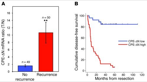
Early this morning I saw a headline float by my Twitter stream from yesterday with a link to an article or paper suggesting that yes, we can indeed predict metastasis. I can’t remember who shared it, or what was the exact news article but a quick Google search for latest news found some noise around a potential biomarker, CPE-ΔN. The paper (open access) in the references link below, is from the Journal of Clinical Investigation.

Now, the idea that a biomarker might be able to predict metastasis is important because it signals the need for more aggressive treatment as the disease is advancing. Equally, if someone is doing well, you don’t want to intensify therapy needlessly but resection may be more appropriate.   Clearly, the earlier you detect the cancer, the better, but conversely, figuring out when to change treatment and prevent or slow metastasis is also important.

Reading the paper carefully, the authors stated:

“We report here that the carboxypeptidase E gene (CPE) is alternatively spliced in human tumors to yield an N-terminal truncated protein (CPE-ΔN) that drives metastasis.”

In the research, they used a liver cancer model (or hepatocellular cancer, HCC) to see what was happening with the protein.  Interestingly, CPE-ΔN tended to be present and have high levels in tumours that have metastasised.

They followed a group of patients with HCC (n=99) and looked to determine whether high or low levels of CPE-ΔN was associated with prognosis, with interesting results:

Can cancer metastasis be predicted?

They also looked at patients with stage 2 disease that had only spread within the liver, as well as patients with a rare adrenal disease and colon cancer.  The patients with stage II HCC have a low chance of recurrence, but it can happen, so the question was could the biomarker be used to predict those most at risk?

The answer was yes.

Of the patients with early HCC (n=18):

  • Thirteen had low levels of CPE-ΔN and 10 of those were still cancer-free three years after surgery.   However, three with low CPE-delta N levels did have recurrence, giving an accuracy level of 77% in predicting metastasis.
  • Five had high levels of CPE-ΔN and in four of them recurrence occurred, giving an accuracy level of 90%.

All in all, it’s a good piece of solid research that may have important implications for future research.  Be warned, the paper is a little heavy to read though!

The next steps for the group are:

  1. Find a therapeutic method of blocking CPE-ΔN, preferably with a small molecule
  2. Determine the mechanism by which CPE-ΔN is activated, thereby figuring out how the switching on of metastasis works

All in all, although this research, while still at the very early stage, looks promising and worth following to see how the idea pans out.  I can’t help wondering how this research will impact the Norton and Massagué cancer seeding theory – it should add to it.  Now, if only we can find out what activates the CPE-ΔN protein, thereby triggering the metastasis, that could well be a key piece in the puzzle.

References:

ResearchBlogging.orgLee, T., Murthy, S., Cawley, N., Dhanvantari, S., Hewitt, S., Lou, H., Lau, T., Ma, S., Huynh, T., Wesley, R., Ng, I., Pacak, K., Poon, R., & Loh, Y. (2011). An N-terminal truncated carboxypeptidase E splice isoform induces tumor growth and is a biomarker for predicting future metastasis in human cancers Journal of Clinical Investigation DOI: 10.1172/JCI40433

“Ubiquitin-dependent mechanisms have emerged as essential regulatory elements controlling cellular levels of Smads and TGFβ-dependent biological outputs such as epithelial–mesenchymal transition (EMT).

In this study, we identify a HECT E3 ubiquitin ligase known as WWP2 (Full-length WWP2-FL), together with two WWP2 isoforms (N-terminal, WWP2-N; C-terminal WWP2-C), as novel Smad-binding partners. We show that WWP2-FL interacts exclusively with Smad2, Smad3 and Smad7 in the TGFβ pathway.

Interestingly, the WWP2-N isoform interacts with Smad2 and Smad3, whereas WWP2-C interacts only with Smad7. In addition, WWP2-FL and WWP2-C have a preference for Smad7 based on protein turnover and ubiquitination studies. Unexpectedly, we also find that WWP2-N, which lacks the HECT ubiquitin ligase domain, can also interact with WWP2-FL in a TGFβ-regulated manner and activate endogenous WWP2 ubiquitin ligase activity causing degradation of unstimulated Smad2 and Smad3.

Consistent with our protein interaction data, overexpression and knockdown approaches reveal that WWP2 isoforms differentially modulate TGFβ-dependent transcription and EMT.

Finally, we show that selective disruption of WWP2 interactions with inhibitory Smad7 can stabilise Smad7 protein levels and prevent TGFβ-induced EMT.

Collectively, our data suggest that WWP2-N can stimulate WWP2-FL leading to increased activity against unstimulated Smad2 and Smad3, and that Smad7 is a preferred substrate for WWP2-FL and WWP2-C following prolonged TGFβ stimulation.

Significantly, this is the first report of an interdependent biological role for distinct HECT E3 ubiquitin ligase isoforms, and highlights an entirely novel regulatory paradigm that selectively limits the level of inhibitory and activating Smads.”

Source: Oncogene

That was an abstract I was browsing over coffee in my oncology RSS feeds and while it was a bit heavy for early in the day, I was intrigued because Smads have been cropping up in GI sessions at meetings over the last six months or so.  Smads are signal transducers for members of the transforming growth factor-beta (TGF-beta) superfamily, so they occupy a key role in transcription of proteins:

The biology of TGF-beta and Smads

In addition, I’ve included a link to an open access article on the biology of Smads in the references below.

Essentially, the translational research from Soond and Chantry (2011) is suggesting that blocking the WWP2 gene could prevent metastasis, ie cancers from spreading to other organs of the body.  Many of you will remember the post on Norton and Massague’s cancer cell seeding theory and this new finding could well have implications for that research too.

Overall, the latest findings mean that if we have a valid target, we can design a drug to target the rogue gene sending signals.

Of course, these are still very early days yet, but it will be interesting to see if the basic science can be translated into R&D and eventually, a real clinical impact in the long run.

For those of you wanting a simpler version of the abstract, BBC Health did a nice job of putting the research into plain English.  Do check out their short report with pretty pictures here.

References:

ResearchBlogging.orgSoond, S., & Chantry, A. (2011). Selective targeting of activating and inhibitory Smads by distinct WWP2 ubiquitin ligase isoforms differentially modulates TGFβ signalling and EMT Oncogene DOI: 10.1038/onc.2010.617

Attisano, L., & Tuen Lee-Hoeflich, S. (2001). The Smads Genome Biology, 2 (8) DOI: 10.1186/gb-2001-2-8-reviews3010

Izzi, L., & Attisano, L. (2004). Regulation of the TGFβ signalling pathway by ubiquitin-mediated degradation Oncogene, 23 (11), 2071-2078 DOI: 10.1038/sj.onc.1207412

That was the question from a reader that greeted me in my inbox recently, it’s a good point.   Sorafenib has received FDA approval in this indication, while Pfizer terminated their phase III trial of sunitinib in HCC for futility last year.

At first, I couldn’t remember the subtle differences between them, since they both inhibit VEGF and PDGF, although sunitinib also inhibits KIT, until a friend reminded that sorafenib also targets RAF.  On checking out the IC50 values of several multi-kinase TKIs, it turned out to be true, good catch:

Why does sorafenib work in HCC but sunitinib doesn't

In addition, there was a paper just published from Nagai et al., (2011) in Molecular Cancer Therapeutics, which demonstrated sorafenib inhibits the hepatocyte growth factor (HGF) mediated epithelial mesenchymal transition (EMT) in hepatocellular carcinoma (HCC).  EMT is a key developmental program that is often activated during cancer invasion and metastasis.  It is a highly complex area that is receiving a lot of research attention at the moment.

Nagai et al., essentially demonstrated that:

“Sorafenib and the MEK inhibitor U0126 markedly inhibited the HGF-induced morphologic changes, SNAI1 upregulation, and cadherin switching, whereas the PI3 kinase inhibitor wortmannin did not.

Collectively, these findings indicate that sorafenib downregulates SNAI1 expression by inhibiting mitogen-activated protein kinase (MAPK) signaling, thereby inhibiting the EMT in HCC cells.”

If we look at the potential pathway, it would look like this:

HGF-RAF-EMT pathway

Based on these findings, it would be interesting to find out whether adding a MEK inhibitor to sorafenib would improve efficacy further to cut off feedback loops.

On checking out the clinical trials database, I found two trials either ongoing or about to begin enrolling with sorafenib and AZD6244 (ARRY 142886), a MEK inhibitor from AstraZeneca/Array Pharma.  The former should have data emerging soon, if it hasn’t already done so.  I think EMT is a fertile area of research where we may see more science and emerging this year.

References:

ResearchBlogging.org Nagai, T., Arao, T., Furuta, K., Sakai, K., Kudo, K., Kaneda, H., Tamura, D., Aomatsu, K., Kimura, H., Fujita, Y., Matsumoto, K., Saijo, N., Kudo, M., & Nishio, K. (2011). Sorafenib Inhibits the Hepatocyte Growth Factor-Mediated Epithelial Mesenchymal Transition in Hepatocellular Carcinoma Molecular Cancer Therapeutics, 10 (1), 169-177 DOI: 10.1158/1535-7163.MCT-10-0544

error: Content is protected !!